1KIX | pdb_00001kix

Dimeric Structure of the O. nova Telomere End Binding Protein Alpha Subunit with Bound ssDNA


Experimental Data Snapshot

  • Method: X-RAY DIFFRACTION
  • Resolution: 2.70 Å
  • R-Value Free: 
    0.240 (Depositor), 0.246 (DCC) 
  • R-Value Work: 
    0.208 (Depositor), 0.216 (DCC) 
  • R-Value Observed: 
    0.208 (Depositor) 

Starting Model: experimental
View more details

wwPDB Validation 3D Report Full Report

Validation slider image for 1KIX

This is version 1.3 of the entry. See complete history

Literature

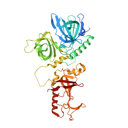
Dimeric structure of the Oxytricha nova telomere end-binding protein alpha-subunit bound to ssDNA.

Peersen, O.B.Ruggles, J.A.Schultz, S.C.

(2002) Nat Struct Biol 9: 182-187

  • DOI: https://doi.org/10.1038/nsb761
  • Primary Citation Related Structures: 
    1KIX

  • PubMed Abstract: 

    Telomeres are the specialized protein--DNA complexes that cap and protect the ends of linear eukaryotic chromosomes. The extreme 3' end of the telomeric DNA in Oxytricha nova is bound by a two-subunit sequence-specific and 3' end-specific protein called the telomere end-binding protein (OnTEBP). Here we describe the crystal structure of the alpha-subunit of OnTEBP in complex with T4G4 single-stranded telomeric DNA. This structure shows an (alpha--ssDNA)2 homodimer with a large approximately 7,000 A2 protein--protein interface in which the domains of alpha are rearranged extensively from their positions in the structure of an alpha--beta--ssDNA ternary complex. The (alpha--ssDNA)2 complex can bind two telomeres on opposite sides of the dimer and, thus, acts as a protein mediator of telomere--telomere associations. The structures of the (alpha--ssDNA)2 dimer presented here and the previously described alpha--beta--ssDNA complex demonstrate that OnTEBP forms multiple telomeric complexes that potentially mediate the assembly and disassembly of higher order telomeric structures.


  • Organizational Affiliation
    • University of Colorado, Department of Chemistry and Biochemistry, Boulder, Colorado 80309-0215 USA. Olve.Peersen@ColoState.edu

Macromolecule Content 

  • Total Structure Weight: 58.85 kDa 
  • Atom Count: 3,837 
  • Modeled Residue Count: 453 
  • Deposited Residue Count: 503 
  • Unique protein chains: 1
  • Unique nucleic acid chains: 1

Macromolecules


Find similar proteins by:|  3D Structure
Entity ID: 2
MoleculeChains  Sequence LengthOrganismDetailsImage
Telomere-Binding Protein alpha SubunitB [auth A]495Sterkiella novaMutation(s): 0 
Gene Names: MAC-56A
UniProt
Find proteins for P29549 (Sterkiella nova)
Explore P29549 
Go to UniProtKB:  P29549
Entity Groups
Sequence Clusters30% Identity50% Identity70% Identity90% Identity95% Identity100% Identity
UniProt GroupP29549
Sequence Annotations
Expand
Reference Sequence
Find similar nucleic acids by:  Sequence
Entity ID: 1
MoleculeChains LengthOrganismImage
5'-D(*T*TP*TP*TP*GP*GP*GP*G)-3'A [auth D]8N/A
Sequence Annotations
Expand
Reference Sequence

Experimental Data & Validation

Experimental Data

  • Method: X-RAY DIFFRACTION
  • Resolution: 2.70 Å
  • R-Value Free:  0.240 (Depositor), 0.246 (DCC) 
  • R-Value Work:  0.208 (Depositor), 0.216 (DCC) 
  • R-Value Observed: 0.208 (Depositor) 
Space Group: F 4 3 2
Unit Cell:
Length ( Å )Angle ( ˚ )
a = 284α = 90
b = 284β = 90
c = 284γ = 90
Software Package:
Software NamePurpose
X-PLORmodel building
CNSrefinement
DENZOdata reduction
SCALEPACKdata scaling
X-PLORphasing

Structure Validation

View Full Validation Report


Entry History 

Deposition Data

Revision History  (Full details and data files)

  • Version 1.0: 2002-02-22
    Type: Initial release
  • Version 1.1: 2008-04-27
    Changes: Version format compliance
  • Version 1.2: 2011-07-13
    Changes: Version format compliance
  • Version 1.3: 2023-08-16
    Changes: Data collection, Database references, Derived calculations, Refinement description