1KCM | pdb_00001kcm

Crystal Structure of Mouse PITP Alpha Void of Bound Phospholipid at 2.0 Angstroms Resolution


Experimental Data Snapshot

  • Method: X-RAY DIFFRACTION
  • Resolution: 2.00 Å
  • R-Value Free: 
    0.273 (Depositor), 0.294 (DCC) 
  • R-Value Work: 
    0.216 (Depositor), 0.244 (DCC) 
  • R-Value Observed: 
    0.219 (Depositor) 

Starting Model: experimental
View more details

wwPDB Validation 3D Report Full Report

Validation slider image for 1KCM

This is version 1.3 of the entry. See complete history

Literature

Structure of apo-phosphatidylinositol transfer protein alpha provides insight into membrane association.

Schouten, A.Agianian, B.Westerman, J.Kroon, J.Wirtz, K.W.Gros, P.

(2002) EMBO J 21: 2117-2121

  • DOI: https://doi.org/10.1093/emboj/21.9.2117
  • Primary Citation Related Structures: 
    1KCM

  • PubMed Abstract: 

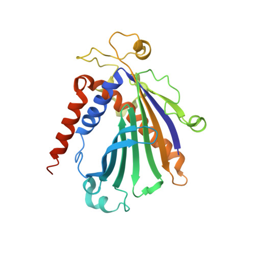
    Phosphatidylinositol transfer protein alpha (PITP alpha) is a ubiquitous and highly conserved protein in multicellular eukaryotes that catalyzes the exchange of phospholipids between membranes in vitro and participates in cellular phospholipid metabolism, signal transduction and vesicular trafficking in vivo. Here we report the three-dimensional crystal structure of a phospholipid-free mouse PITP alpha at 2.0 A resolution. The structure reveals an open conformation characterized by a channel running through the protein. The channel is created by opening the phospholipid-binding cavity on one side by displacement of the C-terminal region and a hydrophobic lipid exchange loop, and on the other side by flattening of the central beta-sheet. The relaxed conformation is stabilized at the proposed membrane association site by hydrophobic interactions with a crystallographically related molecule, creating an intimate dimer. The observed open conformer is consistent with a membrane-bound state of PITP and suggests a mechanism for membrane anchoring and the presentation of phosphatidylinositol to kinases and phospholipases after its extraction from the membrane. Coordinates have been deposited in the Protein Data Bank (accession No. 1KCM).


  • Organizational Affiliation
    • Department of Crystal and Structural Chemistry, Bijvoet Center for Biomolecular Research, Utrecht University, Padualaan 8,NL-3584 CH Utrecht, The Netherlands.

Macromolecule Content 

  • Total Structure Weight: 31.81 kDa 
  • Atom Count: 2,346 
  • Modeled Residue Count: 256 
  • Deposited Residue Count: 270 
  • Unique protein chains: 1

Macromolecules

Find similar proteins by:|  3D Structure
Entity ID: 1
MoleculeChains  Sequence LengthOrganismDetailsImage
Phosphatidylinositol Transfer Protein alpha270Mus musculusMutation(s): 0 
UniProt & NIH Common Fund Data Resources
Find proteins for P53810 (Mus musculus)
Explore P53810 
Go to UniProtKB:  P53810
IMPC:  MGI:99887
Entity Groups
Sequence Clusters30% Identity50% Identity70% Identity90% Identity95% Identity100% Identity
UniProt GroupP53810
Sequence Annotations
Expand
Reference Sequence

Experimental Data & Validation

Experimental Data

  • Method: X-RAY DIFFRACTION
  • Resolution: 2.00 Å
  • R-Value Free:  0.273 (Depositor), 0.294 (DCC) 
  • R-Value Work:  0.216 (Depositor), 0.244 (DCC) 
  • R-Value Observed: 0.219 (Depositor) 
Space Group: P 32 2 1
Unit Cell:
Length ( Å )Angle ( ˚ )
a = 50.464α = 90
b = 50.464β = 90
c = 216.105γ = 120
Software Package:
Software NamePurpose
AMoREphasing
REFMACrefinement
DENZOdata reduction
SCALEPACKdata scaling

Structure Validation

View Full Validation Report


Entry History 

Deposition Data

Revision History  (Full details and data files)

  • Version 1.0: 2002-05-08
    Type: Initial release
  • Version 1.1: 2008-04-27
    Changes: Version format compliance
  • Version 1.2: 2011-07-13
    Changes: Derived calculations, Version format compliance
  • Version 1.3: 2023-08-16
    Changes: Data collection, Database references, Refinement description