1KCG | pdb_00001kcg

NKG2D in complex with ULBP3


Experimental Data Snapshot

  • Method: X-RAY DIFFRACTION
  • Resolution: 2.60 Å
  • R-Value Free: 
    0.268 (Depositor) 
  • R-Value Work: 
    0.220 (Depositor) 

wwPDB Validation   3D Report Full Report


This is version 2.0 of the entry. See complete history


Literature

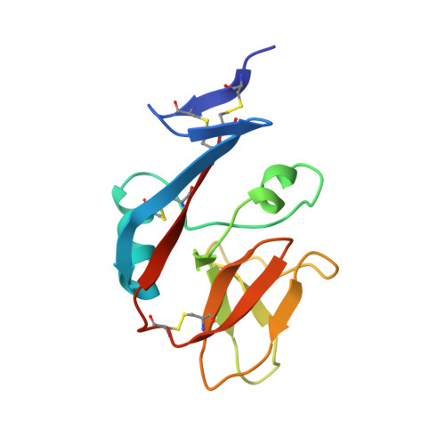
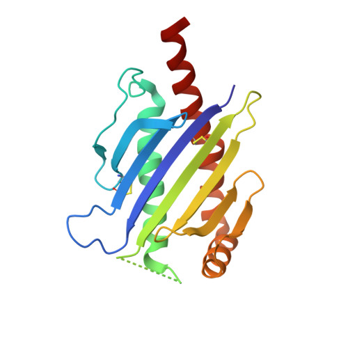
Conformational plasticity revealed by the cocrystal structure of NKG2D and its class I MHC-like ligand ULBP3.

Radaev, S.Rostro, B.Brooks, A.G.Colonna, M.Sun, P.D.

(2001) Immunity 15: 1039-1049

  • DOI: https://doi.org/10.1016/s1074-7613(01)00241-2
  • Primary Citation of Related Structures:  
    1KCG

  • PubMed Abstract: 

    NKG2D is known to trigger the natural killer (NK) cell lysis of various tumor and virally infected cells. In the NKG2D/ULBP3 complex, the structure of ULBP3 resembles the alpha1 and alpha2 domains of classical MHC molecules without a bound peptide. The lack of alpha3 and beta2m domains is compensated by replacing two hydrophobic patches at the underside of the class I MHC-like beta sheet floor with a group of hydrophilic and charged residues in ULBP3. NKG2D binds diagonally across the ULBP3 alpha helices, creating a complementary interface, an asymmetrical subunit orientation, and local conformational adjustments in the receptor. The interface is stabilized primarily by hydrogen bonds and hydrophobic interactions. Unlike the KIR receptors that recognize a conserved HLA region by a lock-and-key mechanism, NKG2D recognizes diverse ligands by an induced-fit mechanism.


  • Organizational Affiliation
    • Structural Biology Section, Laboratory of Immunogenetics, National Institute of Allergy and Infectious Diseases, National Institutes of Health, 12441 Parklawn Drive, Rockville, MD 20852, USA.

Macromolecules
Find similar proteins by:  (by identity cutoff)  |  3D Structure
Entity ID: 1
MoleculeChains Sequence LengthOrganismDetailsImage
NKG2-D type II integral membrane protein
A, B
124Homo sapiensMutation(s): 0 
Gene Names: KLRK1D12S2489ENKG2D
UniProt & NIH Common Fund Data Resources
Find proteins for P26718 (Homo sapiens)
Explore P26718 
Go to UniProtKB:  P26718
PHAROS:  P26718
GTEx:  ENSG00000213809 
Entity Groups  
Sequence Clusters30% Identity50% Identity70% Identity90% Identity95% Identity100% Identity
UniProt GroupP26718
Sequence Annotations
Expand
  • Reference Sequence
Find similar proteins by:  (by identity cutoff)  |  3D Structure
Entity ID: 2
MoleculeChains Sequence LengthOrganismDetailsImage
UL16-binding protein 3178Homo sapiensMutation(s): 0 
Gene Names: ULBP3N2DL3RAET1N
UniProt & NIH Common Fund Data Resources
Find proteins for Q9BZM4 (Homo sapiens)
Explore Q9BZM4 
Go to UniProtKB:  Q9BZM4
PHAROS:  Q9BZM4
GTEx:  ENSG00000131019 
Entity Groups  
Sequence Clusters30% Identity50% Identity70% Identity90% Identity95% Identity100% Identity
UniProt GroupQ9BZM4
Sequence Annotations
Expand
  • Reference Sequence
Small Molecules
Modified Residues  1 Unique
IDChains TypeFormula2D DiagramParent
CGL
Query on CGL
C
L-PEPTIDE LINKINGC13 H22 N4 O8 S2CYS
Experimental Data & Validation

Experimental Data

  • Method: X-RAY DIFFRACTION
  • Resolution: 2.60 Å
  • R-Value Free:  0.268 (Depositor) 
  • R-Value Work:  0.220 (Depositor) 
Space Group: P 43 21 2
Unit Cell:
Length ( Å )Angle ( ˚ )
a = 62.05α = 90
b = 62.05β = 90
c = 237.31γ = 90
Software Package:
Software NamePurpose
MAR345data collection
HKL-2000data reduction
SOLVEphasing
CNSrefinement
HKL-2000data scaling

Structure Validation

View Full Validation Report


Entry History 

Deposition Data

Revision History  (Full details and data files)

  • Version 1.0: 2002-01-09
    Type: Initial release
  • Version 1.1: 2008-04-27
    Changes: Version format compliance
  • Version 1.2: 2011-07-13
    Changes: Version format compliance
  • Version 2.0: 2024-12-25
    Changes: Atomic model, Data collection, Database references, Derived calculations, Non-polymer description, Polymer sequence, Refinement description, Source and taxonomy, Structure summary