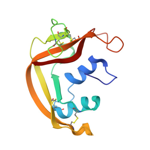
1K2A

Modified Form of Eosinophil-derived Neurotoxin


Experimental Data Snapshot

  • Method: X-RAY DIFFRACTION
  • Resolution: 1.00 Å
  • R-Value Free: 0.169 
  • R-Value Work: 0.132 
  • R-Value Observed: 0.135 

wwPDB Validation   3D Report Full Report


This is version 1.3 of the entry. See complete history


Literature

Crystallographic and functional studies of a modified form of eosinophil-derived neurotoxin (EDN) with novel biological activities.

Chang, C.Newton, D.L.Rybak, S.M.Wlodawer, A.

(2002) J Mol Biol 317: 119-130

  • DOI: https://doi.org/10.1006/jmbi.2002.5406
  • Primary Citation of Related Structures:  
    1K2A

  • PubMed Abstract: 

    The crystal structure of a post-translationally modified form of eosinophil-derived neurotoxin (EDN) with four extra residues on its N terminus ((-4)EDN) has been solved and refined at atomic resolution (1 A). Two of the extra residues can be placed unambiguously, while the density corresponding to two others is poor. The modified N terminus appears to influence the position of the catalytically important His129, possibly explaining the diminished catalytic activity of this variant. However, (-4)EDN has been shown to be cytotoxic to a Kaposi's sarcoma tumor cell line and other endothelial cell lines. Analysis of the structure and function suggests that the reason for cytotoxicity is most likely due to cellular recognition by the N-terminal extension, since the intrinsic activity of the enzyme is not sufficient for cytotoxicity and the N-terminal extension does not affect the conformation of EDN.


  • Organizational Affiliation

    Macromolecular Crystallography Laboratory, National Cancer Institute, Frederick, MD 21702, USA.


Macromolecules
Find similar proteins by:  (by identity cutoff)  |  3D Structure
Entity ID: 1
MoleculeChains Sequence LengthOrganismDetailsImage
eosinophil-derived neurotoxin136Homo sapiensMutation(s): 0 
EC: 3.1.27.5 (PDB Primary Data), 4.6.1.18 (UniProt)
UniProt & NIH Common Fund Data Resources
Find proteins for P10153 (Homo sapiens)
Explore P10153 
Go to UniProtKB:  P10153
PHAROS:  P10153
GTEx:  ENSG00000169385 
Entity Groups  
Sequence Clusters30% Identity50% Identity70% Identity90% Identity95% Identity100% Identity
UniProt GroupP10153
Sequence Annotations
Expand
  • Reference Sequence
Experimental Data & Validation

Experimental Data

  • Method: X-RAY DIFFRACTION
  • Resolution: 1.00 Å
  • R-Value Free: 0.169 
  • R-Value Work: 0.132 
  • R-Value Observed: 0.135 
  • Space Group: P 21 21 21
Unit Cell:
Length ( Å )Angle ( ˚ )
a = 42.138α = 90
b = 52.558β = 90
c = 56.627γ = 90
Software Package:
Software NamePurpose
DENZOdata reduction
SCALEPACKdata scaling
AMoREphasing
SHELXL-97refinement

Structure Validation

View Full Validation Report


Entry History 

Deposition Data

Revision History  (Full details and data files)

  • Version 1.0: 2002-04-03
    Type: Initial release
  • Version 1.1: 2008-04-27
    Changes: Version format compliance
  • Version 1.2: 2011-07-13
    Changes: Version format compliance
  • Version 1.3: 2024-11-06
    Changes: Data collection, Database references, Derived calculations, Structure summary