1JUY | pdb_00001juy

REFINED CRYSTAL STRUCTURE OF ADENYLOSUCCINATE SYNTHETASE FROM ESCHERICHIA COLI COMPLEXED WITH HYDANTOCIDIN 5'-PHOSPHATE GDP, HPO4(2-), MG2+, AND HADACIDIN


Experimental Data Snapshot

  • Method: X-RAY DIFFRACTION
  • Resolution: 2.50 Å
  • R-Value Free: 
    0.250 (Depositor) 
  • R-Value Work: 
    0.195 (Depositor), 0.211 (DCC) 
  • R-Value Observed: 
    0.195 (Depositor) 

wwPDB Validation 3D Report Full Report

Validation slider image for 1JUY

Ligand Structure Quality Assessment 


This is version 1.4 of the entry. See complete history

Literature

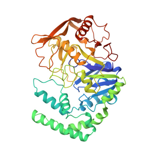
Refined crystal structure of adenylosuccinate synthetase from Escherichia coli complexed with hydantocidin 5'-phosphate, GDP, HPO4(2-), Mg2+, and hadacidin.

Poland, B.W.Lee, S.F.Subramanian, M.V.Siehl, D.L.Anderson, R.J.Fromm, H.J.Honzatko, R.B.

(1996) Biochemistry 35: 15753-15759

  • DOI: https://doi.org/10.1021/bi961758r
  • Primary Citation Related Structures: 
    1JUY

  • PubMed Abstract: 

    A crystal structure of adenylosuccinate synthetase from Escherichia coli, complexed with 5'-phosphate, GDP, HPO4(2-), Mg2+, and hadacidin at 100 K, has been refined to an Rfactor of 0.195 against data to 2.6 A resolution. Bond lengths and angles deviate from expected values by 0.012 A and 1.86 degrees, respectively. Lys 16 and backbone amides 15-17 and 42 interact with the phosphates of GDP, while Ser 414, Asp 333, and backbone amides 331 and 416 interact with the base. Mg2+ is octahedrally coordinated. Oxygen atoms from GDP, phosphate, and hadacidin define the equatorial plane of coordination of the Mg2+, while backbone carbonyl 40 and the side chain of Asp 13 are the apical ligands. HPO4(2-) hydrogen bonds with Lys 16, His 41, backbone amides 13, 40, and 224, and the base moiety of the hydantocidin inhibitor. The carboxylate of hadacidin interacts with Arg 303 and Thr 301; its N-formyl group coordinates to Mg2+, and its hydroxyl group hydrogen bonds with Asp 13. The 5'-phosphate of the hydantocidin inhibitor interacts with Asn 38, Thr 129, and Thr 239 but is approximately 3.5 A from Arg 143 (related by molecular 2-fold symmetry). The base moiety of hydantocidin 5'-phosphate hydrogen bonds to Gln 224 and participates in a hydrogen-bonded network that includes the phosphate molecule, several water molecules, and Asp 13. Hydantocidin 5'-phosphate, GDP, HPO4(2-), and Mg2+ may represent a set of synergistic inhibitors even more effective than the combination of IMP, GDP, NO3-, and Mg2+.


  • Organizational Affiliation
    • Department of Biochemistry and Biophysics, Iowa State University, Ames 50011, USA.

Macromolecule Content 

  • Total Structure Weight: 48.25 kDa 
  • Atom Count: 3,615 
  • Modeled Residue Count: 431 
  • Deposited Residue Count: 431 
  • Unique protein chains: 1

Macromolecules

Find similar proteins by:|  3D Structure
Entity ID: 1
MoleculeChains  Sequence LengthOrganismDetailsImage
ADENYLOSUCCINATE SYNTHETASE431Escherichia coliMutation(s): 0 
EC: 6.3.4.4
UniProt
Find proteins for P0A7D4 (Escherichia coli (strain K12))
Explore P0A7D4 
Go to UniProtKB:  P0A7D4
Entity Groups
Sequence Clusters30% Identity50% Identity70% Identity90% Identity95% Identity100% Identity
UniProt GroupP0A7D4
Sequence Annotations
Expand
Reference Sequence

Small Molecules

Ligands 5 Unique
IDChains Name / Formula / InChI Key2D Diagram3D Interactions
GDP

Query on GDP


Download:Ideal Coordinates CCD File
D [auth A]GUANOSINE-5'-DIPHOSPHATE
C10 H15 N5 O11 P2
QGWNDRXFNXRZMB-UUOKFMHZSA-N
H5P

Query on H5P


Download:Ideal Coordinates CCD File
E [auth A]HYDANTOCIDIN-5'-PHOSPHATE
C7 H11 N2 O9 P
HVXIMXHBUJADCC-GTBMBKLPSA-N
HDA

Query on HDA


Download:Ideal Coordinates CCD File
F [auth A]HADACIDIN
C3 H5 N O4
URJHVPKUWOUENU-UHFFFAOYSA-N
PI

Query on PI


Download:Ideal Coordinates CCD File
C [auth A]HYDROGENPHOSPHATE ION
H O4 P
NBIIXXVUZAFLBC-UHFFFAOYSA-L
MG

Query on MG


Download:Ideal Coordinates CCD File
B [auth A]MAGNESIUM ION
Mg
JLVVSXFLKOJNIY-UHFFFAOYSA-N

Experimental Data & Validation

Experimental Data

  • Method: X-RAY DIFFRACTION
  • Resolution: 2.50 Å
  • R-Value Free:  0.250 (Depositor) 
  • R-Value Work:  0.195 (Depositor), 0.211 (DCC) 
  • R-Value Observed: 0.195 (Depositor) 
Space Group: P 32 2 1
Unit Cell:
Length ( Å )Angle ( ˚ )
a = 80.4α = 90
b = 80.4β = 90
c = 158.41γ = 120
Software Package:
Software NamePurpose
X-PLORmodel building
X-PLORrefinement
XENGENdata reduction
XENGENdata scaling
X-PLORphasing

Structure Validation

View Full Validation Report


Ligand Structure Quality Assessment 


Entry History 

Deposition Data

Revision History  (Full details and data files)

  • Version 1.0: 1997-06-24
    Type: Initial release
  • Version 1.1: 2008-03-27
    Changes: Version format compliance
  • Version 1.2: 2011-07-13
    Changes: Version format compliance
  • Version 1.3: 2017-11-29
    Changes: Derived calculations, Other
  • Version 1.4: 2024-02-07
    Changes: Data collection, Database references, Derived calculations