1JRR

HUMAN PLASMINOGEN ACTIVATOR INHIBITOR-2.[LOOP (66-98) DELETIONMUTANT] COMPLEXED WITH PEPTIDE MIMIckING THE REACTIVE CENTER LOOP


Experimental Data Snapshot

  • Method: X-RAY DIFFRACTION
  • Resolution: 1.60 Å
  • R-Value Free: 0.225 
  • R-Value Work: 0.197 
  • R-Value Observed: 0.198 

wwPDB Validation   3D Report Full Report


This is version 1.2 of the entry. See complete history


Literature

CRYSTAL STRUCTURE OF THE COMPLEX OF PLASMINOGEN ACTIVATOR INHIBITOR 2 WITH A PEPTIDE MIMICKING THE REACTIVE CENTER LOOP

JANKOVA, L.HARROP, S.J.SAUNDERS, D.N.ANDREWS, J.L.BERTRAM, K.C.GOULD, A.R.BAKER, M.S.CURMI, P.M.G.

(2001) J Biol Chem 276: 43374-43382

  • DOI: https://doi.org/10.1074/jbc.M103021200
  • Primary Citation of Related Structures:  
    1JRR

  • PubMed Abstract: 

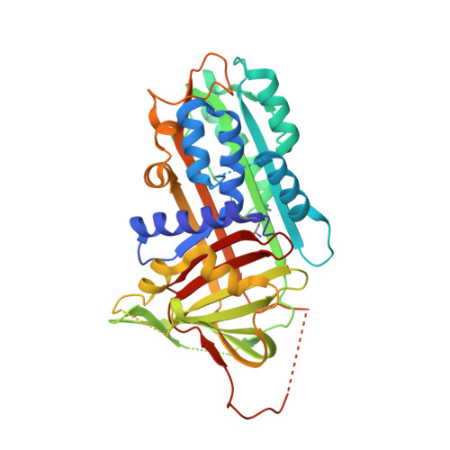
    The structure of the serpin, plasminogen activator inhibitor type-2 (PAI-2), in a complex with a peptide mimicking its reactive center loop (RCL) has been determined at 1.6-A resolution. The structure shows the relaxed state serpin structure with a prominent six-stranded beta-sheet. Clear electron density is seen for all residues in the peptide. The P1 residue of the peptide binds to a well defined pocket at the base of PAI-2 that may be important in determining the specificity of protease inhibition. The stressed-to-relaxed state (S --> R) transition in PAI-2 can be modeled as the relative motion between a quasirigid core domain and a smaller segment comprising helix hF and beta-strands s1A, s2A, and s3A. A comparison of the Ramachandran plots of the stressed and relaxed state PAI-2 structures reveals the location of several hinge regions connecting these two domains. The hinge regions cluster in three locations on the structure, ensuring a cooperative S --> R transition. We hypothesize that the hinge formed by the conserved Gly(206) on beta-strand s3A in the breach region of PAI-2 effects the S --> R transition by altering its backbone torsion angles. This torsional change is due to the binding of the P14 threonine of the RCL to the open breach region of PAI-2.


  • Organizational Affiliation

    Initiative in Biomolecular Structure, School of Physics, University of New South Wales, Sydney New South Wales 2052, Australia.


Macromolecules
Find similar proteins by:  (by identity cutoff)  |  3D Structure
Entity ID: 1
MoleculeChains Sequence LengthOrganismDetailsImage
PLASMINOGEN ACTIVATOR INHIBITOR-2382Homo sapiensMutation(s): 0 
UniProt & NIH Common Fund Data Resources
Find proteins for P05120 (Homo sapiens)
Explore P05120 
Go to UniProtKB:  P05120
PHAROS:  P05120
GTEx:  ENSG00000197632 
Entity Groups  
Sequence Clusters30% Identity50% Identity70% Identity90% Identity95% Identity100% Identity
UniProt GroupP05120
Sequence Annotations
Expand
  • Reference Sequence

Find similar proteins by:  Sequence   |   3D Structure  

Entity ID: 2
MoleculeChains Sequence LengthOrganismDetailsImage
PLASMINOGEN ACTIVATOR INHIBITOR-2B [auth P]15N/AMutation(s): 1 
UniProt & NIH Common Fund Data Resources
Find proteins for P05120 (Homo sapiens)
Explore P05120 
Go to UniProtKB:  P05120
PHAROS:  P05120
GTEx:  ENSG00000197632 
Entity Groups  
Sequence Clusters30% Identity50% Identity70% Identity90% Identity95% Identity100% Identity
UniProt GroupP05120
Sequence Annotations
Expand
  • Reference Sequence
Small Molecules
Ligands 1 Unique
IDChains Name / Formula / InChI Key2D Diagram3D Interactions
BME
Query on BME

Download Ideal Coordinates CCD File 
C [auth A]BETA-MERCAPTOETHANOL
C2 H6 O S
DGVVWUTYPXICAM-UHFFFAOYSA-N
Experimental Data & Validation

Experimental Data

  • Method: X-RAY DIFFRACTION
  • Resolution: 1.60 Å
  • R-Value Free: 0.225 
  • R-Value Work: 0.197 
  • R-Value Observed: 0.198 
  • Space Group: P 21 21 2
Unit Cell:
Length ( Å )Angle ( ˚ )
a = 93.139α = 90
b = 104.16β = 90
c = 41.395γ = 90
Software Package:
Software NamePurpose
MOSFLMdata reduction
SCALAdata scaling
AMoREphasing
REFMACrefinement
CCP4data scaling

Structure Validation

View Full Validation Report


Entry History 

Deposition Data

Revision History  (Full details and data files)

  • Version 1.0: 2001-09-26
    Type: Initial release
  • Version 1.1: 2008-04-27
    Changes: Version format compliance
  • Version 1.2: 2011-07-13
    Changes: Version format compliance