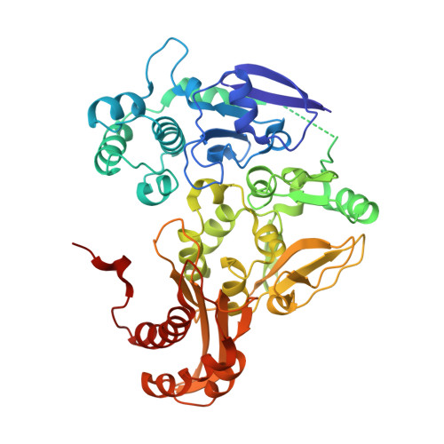
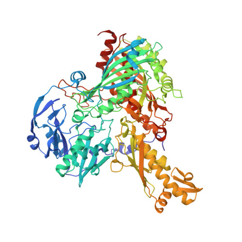
1JRO

Crystal Structure of Xanthine Dehydrogenase from Rhodobacter capsulatus


Experimental Data Snapshot

  • Method: X-RAY DIFFRACTION
  • Resolution: 2.70 Å
  • R-Value Free: 0.252 
  • R-Value Work: 0.213 
  • R-Value Observed: 0.215 

wwPDB Validation   3D Report Full Report


This is version 1.3 of the entry. See complete history


Literature

Crystal structures of the active and alloxanthine-inhibited forms of xanthine dehydrogenase from Rhodobacter capsulatus

Truglio, J.J.Theis, K.Leimkuhler, S.Rappa, R.Rajagopalan, K.V.Kisker, C.

(2002) Structure 10: 115-125

  • DOI: https://doi.org/10.1016/s0969-2126(01)00697-9
  • Primary Citation of Related Structures:  
    1JRO, 1JRP

  • PubMed Abstract: 
  • Xanthine dehydrogenase (XDH), a complex molybdo/iron-sulfur/flavoprotein, catalyzes the oxidation of hypoxanthine to xanthine followed by oxidation of xanthine to uric acid with concomitant reduction of NAD+. The 2.7 A resolution structure of Rhodobacter capsulatus XDH reveals that the bacterial and bovine XDH have highly similar folds despite differences in subunit composition ...

    Xanthine dehydrogenase (XDH), a complex molybdo/iron-sulfur/flavoprotein, catalyzes the oxidation of hypoxanthine to xanthine followed by oxidation of xanthine to uric acid with concomitant reduction of NAD+. The 2.7 A resolution structure of Rhodobacter capsulatus XDH reveals that the bacterial and bovine XDH have highly similar folds despite differences in subunit composition. The NAD+ binding pocket of the bacterial XDH resembles that of the dehydrogenase form of the bovine enzyme rather than that of the oxidase form, which reduces O(2) instead of NAD+. The drug allopurinol is used to treat XDH-catalyzed uric acid build-up occurring in gout or during cancer chemotherapy. As a hypoxanthine analog, it is oxidized to alloxanthine, which cannot be further oxidized but acts as a tight binding inhibitor of XDH. The 3.0 A resolution structure of the XDH-alloxanthine complex shows direct coordination of alloxanthine to the molybdenum via a nitrogen atom. These results provide a starting point for the rational design of new XDH inhibitors.


    Organizational Affiliation

    Department of Pharmacological Sciences, Center for Structural Biology, State University of New York at Stony Brook, Stony Brook, NY 11794, USA.


Macromolecules
Find similar proteins by:  (by identity cutoff)  |  3D Structure
Entity ID: 1
MoleculeChainsSequence LengthOrganismDetailsImage
xanthine dehydrogenase, chain AA, C, E, G462Rhodobacter capsulatusMutation(s): 0 
Gene Names: xdhA
EC: 1.1.1.204
UniProt
Find proteins for O54050 (Rhodobacter capsulatus)
Explore O54050 
Go to UniProtKB:  O54050
Entity Groups  
Sequence Clusters30% Identity50% Identity70% Identity90% Identity95% Identity100% Identity
UniProt GroupO54050
Protein Feature View
Expand
  • Reference Sequence
Find similar proteins by:  (by identity cutoff)  |  3D Structure
Entity ID: 2
MoleculeChainsSequence LengthOrganismDetailsImage
xanthine dehydrogenase, chain BB, D, F, H777Rhodobacter capsulatusMutation(s): 0 
Gene Names: xdhB
EC: 1.1.1.204
UniProt
Find proteins for O54051 (Rhodobacter capsulatus)
Explore O54051 
Go to UniProtKB:  O54051
Entity Groups  
Sequence Clusters30% Identity50% Identity70% Identity90% Identity95% Identity100% Identity
UniProt GroupO54051
Protein Feature View
Expand
  • Reference Sequence
Small Molecules
Ligands 5 Unique
IDChainsName / Formula / InChI Key2D Diagram3D Interactions
FAD
Query on FAD

Download Ideal Coordinates CCD File 
CA [auth G],
K [auth A],
Q [auth C],
W [auth E]
FLAVIN-ADENINE DINUCLEOTIDE
C27 H33 N9 O15 P2
VWWQXMAJTJZDQX-UYBVJOGSSA-N
 Ligand Interaction
MTE
Query on MTE

Download Ideal Coordinates CCD File 
EA [auth H],
M [auth B],
S [auth D],
Y [auth F]
PHOSPHONIC ACIDMONO-(2-AMINO-5,6-DIMERCAPTO-4-OXO-3,7,8A,9,10,10A-HEXAHYDRO-4H-8-OXA-1,3,9,10-TETRAAZA-ANTHRACEN-7-YLMETHYL)ESTER
C10 H14 N5 O6 P S2
HPEUEJRPDGMIMY-IFQPEPLCSA-N
 Ligand Interaction
FES
Query on FES

Download Ideal Coordinates CCD File 
AA [auth G],
BA [auth G],
I [auth A],
J [auth A],
O [auth C],
AA [auth G],
BA [auth G],
I [auth A],
J [auth A],
O [auth C],
P [auth C],
U [auth E],
V [auth E]
FE2/S2 (INORGANIC) CLUSTER
Fe2 S2
NIXDOXVAJZFRNF-UHFFFAOYSA-N
 Ligand Interaction
MOS
Query on MOS

Download Ideal Coordinates CCD File 
FA [auth H],
N [auth B],
T [auth D],
Z [auth F]
DIOXOTHIOMOLYBDENUM(VI) ION
H Mo O2 S
BDSRWPHSAKXXRG-UHFFFAOYSA-M
 Ligand Interaction
CA
Query on CA

Download Ideal Coordinates CCD File 
DA [auth H],
L [auth B],
R [auth D],
X [auth F]
CALCIUM ION
Ca
BHPQYMZQTOCNFJ-UHFFFAOYSA-N
 Ligand Interaction
Experimental Data & Validation

Experimental Data

  • Method: X-RAY DIFFRACTION
  • Resolution: 2.70 Å
  • R-Value Free: 0.252 
  • R-Value Work: 0.213 
  • R-Value Observed: 0.215 
  • Space Group: P 1
Unit Cell:
Length ( Å )Angle ( ˚ )
a = 92.875α = 109.53
b = 141.053β = 105.83
c = 158.113γ = 101.33
Software Package:
Software NamePurpose
DENZOdata reduction
SCALEPACKdata scaling
AMoREphasing
REFMACrefinement

Structure Validation

View Full Validation Report


Entry History 

Deposition Data

Revision History  (Full details and data files)

  • Version 1.0: 2002-01-11
    Type: Initial release
  • Version 1.1: 2008-04-27
    Changes: Version format compliance
  • Version 1.2: 2011-07-13
    Changes: Version format compliance
  • Version 1.3: 2015-04-22
    Changes: Non-polymer description