1JRG

Crystal Structure of the R3 form of Pectate Lyase A, Erwinia chrysanthemi


Experimental Data Snapshot

  • Method: X-RAY DIFFRACTION
  • Resolution: 2.10 Å
  • R-Value Free: 0.211 
  • R-Value Work: 0.160 
  • R-Value Observed: 0.160 

wwPDB Validation   3D Report Full Report


This is version 1.3 of the entry. See complete history


Literature

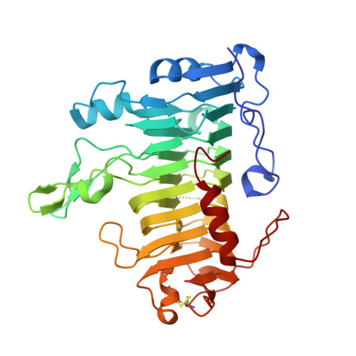
Structure of pectate lyase A: comparison to other isoforms.

Thomas, L.M.Doan, C.N.Oliver, R.L.Yoder, M.D.

(2002) Acta Crystallogr D Biol Crystallogr 58: 1008-1015

  • DOI: https://doi.org/10.1107/s0907444902005851
  • Primary Citation of Related Structures:  
    1JRG, 1JTA

  • PubMed Abstract: 

    Pectate lyase A is a virulence factor secreted by the plant-pathogenic bacteria Erwinia chrysanthemi. The enzyme cleaves the glycosidic bond of pectate polymers by a calcium-dependent beta-elimination mechanism. The crystal structure of pectate lyase A from E. chrysanthemi EC16 has been determined in two crystal forms, monoclinic C2 to 1.8 A and rhombohedral R3 to 2.1 A. The protein structure is compared with two other pectate lyase isoforms from E. chrysanthemi EC16, pectate lyase C and pectate lyase E. Pectate lyase A is unique as it is the only acidic pectate lyase and has end products that are significantly more varied in length in comparison to those of the other four major pectate lyase isozymes. Differences and similarities in polypeptide trace, size and volume of the active-site groove and surface electrostatics are discussed.


  • Organizational Affiliation

    School of Biological Sciences, University of Missouri-Kansas City, 5007 Rockhill Road, Kansas City, MO 64110-2499, USA.


Macromolecules
Find similar proteins by:  (by identity cutoff)  |  3D Structure
Entity ID: 1
MoleculeChains Sequence LengthOrganismDetailsImage
Pectate lyase
A, B
361Dickeya chrysanthemiMutation(s): 0 
Gene Names: PELA
EC: 4.2.2.2
UniProt
Find proteins for P0C1A2 (Dickeya chrysanthemi)
Explore P0C1A2 
Go to UniProtKB:  P0C1A2
Entity Groups  
Sequence Clusters30% Identity50% Identity70% Identity90% Identity95% Identity100% Identity
UniProt GroupP0C1A2
Sequence Annotations
Expand
  • Reference Sequence
Experimental Data & Validation

Experimental Data

  • Method: X-RAY DIFFRACTION
  • Resolution: 2.10 Å
  • R-Value Free: 0.211 
  • R-Value Work: 0.160 
  • R-Value Observed: 0.160 
  • Space Group: H 3
Unit Cell:
Length ( Å )Angle ( ˚ )
a = 98.055α = 90
b = 98.055β = 90
c = 217.162γ = 120
Software Package:
Software NamePurpose
DENZOdata reduction
SCALEPACKdata scaling
AMoREphasing
CNSrefinement

Structure Validation

View Full Validation Report


Entry History 

Deposition Data

Revision History  (Full details and data files)

  • Version 1.0: 2002-06-19
    Type: Initial release
  • Version 1.1: 2008-04-27
    Changes: Version format compliance
  • Version 1.2: 2011-07-13
    Changes: Version format compliance
  • Version 1.3: 2018-04-04
    Changes: Data collection