1JR2 | pdb_00001jr2

Structure of Uroporphyrinogen III Synthase


Experimental Data Snapshot

  • Method: X-RAY DIFFRACTION
  • Resolution: 1.84 Å
  • R-Value Free: 
    0.251 (Depositor), 0.259 (DCC) 
  • R-Value Work: 
    0.200 (Depositor), 0.206 (DCC) 
  • R-Value Observed: 
    0.203 (Depositor) 

wwPDB Validation 3D Report Full Report

Validation slider image for 1JR2

This is version 1.4 of the entry. See complete history

Literature

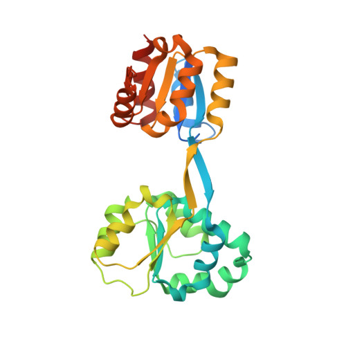
Crystal structure of human uroporphyrinogen III synthase.

Mathews, M.A.Schubert, H.L.Whitby, F.G.Alexander, K.J.Schadick, K.Bergonia, H.A.Phillips, J.D.Hill, C.P.

(2001) EMBO J 20: 5832-5839

  • DOI: https://doi.org/10.1093/emboj/20.21.5832
  • Primary Citation Related Structures: 
    1JR2

  • PubMed Abstract: 

    Uroporphyrinogen III synthase, U3S, the fourth enzyme in the porphyrin biosynthetic pathway, catalyzes cyclization of the linear tetrapyrrole, hydroxymethylbilane, to the macrocyclic uroporphyrino gen III, which is used in several different pathways to form heme, siroheme, chlorophyll, F(430) and vitamin B(12). U3S activity is essential in all organisms, and decreased activity in humans leads to the autosomal recessive disorder congenital erythropoetic porphyria. We have determined the crystal structure of recombinant human U3S at 1.85 A resolution. The protein folds into two alpha/beta domains connected by a beta-ladder. The active site appears to be located between the domains, and variations in relative domain positions observed between crystallographically independent molecules indicates the presence of flexibility that may be important in the catalytic cycle. Possible mechanisms of catalysis were probed by mutating each of the four invariant residues in the protein that have titratable side chains. Additionally, six other highly conserved and titratable side chains were also mutated. In no case, however, did one of these mutations abolish enzyme activity, suggesting that the mechanism does not require acid/base catalysis.


  • Organizational Affiliation
    • Department of Biochemistry, University of Utah School of Medicine, 50 N. Medical Drive, Salt Lake City, UT 84132, USA.

Macromolecule Content 

  • Total Structure Weight: 62.38 kDa 
  • Atom Count: 4,537 
  • Modeled Residue Count: 520 
  • Deposited Residue Count: 572 
  • Unique protein chains: 1

Macromolecules

Find similar proteins by:|  3D Structure
Entity ID: 1
MoleculeChains  Sequence LengthOrganismDetailsImage
UROPORPHYRINOGEN-III SYNTHASE
A, B
286Homo sapiensMutation(s): 0 
Gene Names: UROS
EC: 4.2.1.75
UniProt & NIH Common Fund Data Resources
Find proteins for P10746 (Homo sapiens)
Explore P10746 
Go to UniProtKB:  P10746
PHAROS:  P10746
GTEx:  ENSG00000188690 
Entity Groups
Sequence Clusters30% Identity50% Identity70% Identity90% Identity95% Identity100% Identity
UniProt GroupP10746
Sequence Annotations
Expand
Reference Sequence

Experimental Data & Validation

Experimental Data

  • Method: X-RAY DIFFRACTION
  • Resolution: 1.84 Å
  • R-Value Free:  0.251 (Depositor), 0.259 (DCC) 
  • R-Value Work:  0.200 (Depositor), 0.206 (DCC) 
  • R-Value Observed: 0.203 (Depositor) 
Space Group: P 1
Unit Cell:
Length ( Å )Angle ( ˚ )
a = 39.852α = 80.47
b = 59.251β = 73.32
c = 61.944γ = 88.33
Software Package:
Software NamePurpose
SOLVEphasing
SHARPphasing
REFMACrefinement
DENZOdata reduction
SCALEPACKdata scaling

Structure Validation

View Full Validation Report


Entry History 

Deposition Data

Revision History  (Full details and data files)

  • Version 1.0: 2001-11-07
    Type: Initial release
  • Version 1.1: 2008-04-27
    Changes: Version format compliance
  • Version 1.2: 2011-07-13
    Changes: Version format compliance
  • Version 1.3: 2017-10-04
    Changes: Advisory, Refinement description
  • Version 1.4: 2024-02-07
    Changes: Advisory, Data collection, Database references