1JPP

The Structure of a beta-Catenin Binding Repeat from Adenomatous Polyposis Coli (APC) in Complex with beta-Catenin


Experimental Data Snapshot

  • Method: X-RAY DIFFRACTION
  • Resolution: 3.10 Å
  • R-Value Free: 0.274 
  • R-Value Work: 0.234 
  • R-Value Observed: 0.234 

wwPDB Validation   3D Report Full Report


This is version 1.3 of the entry. See complete history


Literature

Molecular mechanisms of beta-catenin recognition by adenomatous polyposis coli revealed by the structure of an APC-beta-catenin complex.

Eklof Spink, K.Fridman, S.G.Weis, W.I.

(2001) EMBO J 20: 6203-6212

  • DOI: https://doi.org/10.1093/emboj/20.22.6203
  • Primary Citation of Related Structures:  
    1JPP

  • PubMed Abstract: 

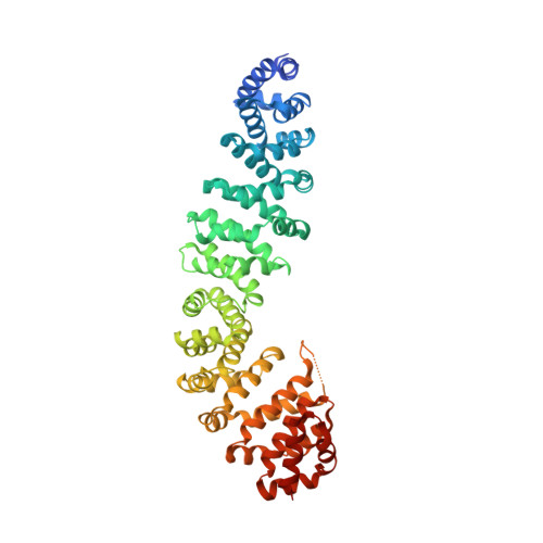
    The adenomatous polyposis coli (APC) tumor suppressor protein plays a critical role in regulating cellular levels of the oncogene product beta-catenin. APC binds to beta-catenin through a series of homologous 15 and 20 amino acid repeats. We have determined the crystal structure of a 15 amino acid beta-catenin binding repeat from APC bound to the armadillo repeat region of beta-catenin. Although it lacks significant sequence homology, the N-terminal half of the repeat binds in a manner similar to portions of E-cadherin and XTcf3, but the remaining interactions are unique to APC. We discuss the implications of this new structure for the design of therapeutics, and present evidence from structural, biochemical and sequence data, which suggest that the 20 amino acid repeats can adopt two modes of binding to beta-catenin.


  • Organizational Affiliation

    Departments of Structural Biology and of Molecular and Cellular Physiology, Stanford University School of Medicine, 299 Campus Dr. West Stanford, CA 94305, USA.


Macromolecules
Find similar proteins by:  (by identity cutoff)  |  3D Structure
Entity ID: 1
MoleculeChains Sequence LengthOrganismDetailsImage
BETA-CATENIN
A, B
538Mus musculusMutation(s): 0 
UniProt & NIH Common Fund Data Resources
Find proteins for Q02248 (Mus musculus)
Explore Q02248 
Go to UniProtKB:  Q02248
IMPC:  MGI:88276
Entity Groups  
Sequence Clusters30% Identity50% Identity70% Identity90% Identity95% Identity100% Identity
UniProt GroupQ02248
Sequence Annotations
Expand
  • Reference Sequence

Find similar proteins by:  Sequence   |   3D Structure  

Entity ID: 2
MoleculeChains Sequence LengthOrganismDetailsImage
ADENOMATOUS POLYPOSIS COLI PROTEIN
C, D
15N/AMutation(s): 0 
UniProt & NIH Common Fund Data Resources
Find proteins for P25054 (Homo sapiens)
Explore P25054 
Go to UniProtKB:  P25054
PHAROS:  P25054
GTEx:  ENSG00000134982 
Entity Groups  
Sequence Clusters30% Identity50% Identity70% Identity90% Identity95% Identity100% Identity
UniProt GroupP25054
Sequence Annotations
Expand
  • Reference Sequence
Small Molecules
Ligands 1 Unique
IDChains Name / Formula / InChI Key2D Diagram3D Interactions
GOL
Query on GOL

Download Ideal Coordinates CCD File 
E [auth B]GLYCEROL
C3 H8 O3
PEDCQBHIVMGVHV-UHFFFAOYSA-N
Experimental Data & Validation

Experimental Data

  • Method: X-RAY DIFFRACTION
  • Resolution: 3.10 Å
  • R-Value Free: 0.274 
  • R-Value Work: 0.234 
  • R-Value Observed: 0.234 
  • Space Group: P 1
Unit Cell:
Length ( Å )Angle ( ˚ )
a = 51.09α = 68.7
b = 69.255β = 87.04
c = 95.099γ = 85.32
Software Package:
Software NamePurpose
CNSrefinement
DENZOdata reduction
SCALEPACKdata scaling
CNSphasing

Structure Validation

View Full Validation Report


Entry History 

Deposition Data

Revision History  (Full details and data files)

  • Version 1.0: 2002-01-16
    Type: Initial release
  • Version 1.1: 2008-04-27
    Changes: Version format compliance
  • Version 1.2: 2011-07-13
    Changes: Non-polymer description, Version format compliance
  • Version 1.3: 2023-08-16
    Changes: Data collection, Database references, Derived calculations, Refinement description