The 2.0 A structure of a cross-linked complex between snowdrop lectin and a branched mannopentaose: evidence for two unique binding modes.
Wright, C.S., Hester, G.(1996) Structure 4: 1339-1352
- PubMed: 8939757 Search on PubMed
- DOI: https://doi.org/10.1016/s0969-2126(96)00141-4
- Primary Citation Related Structures:
1JPC - PubMed Abstract:
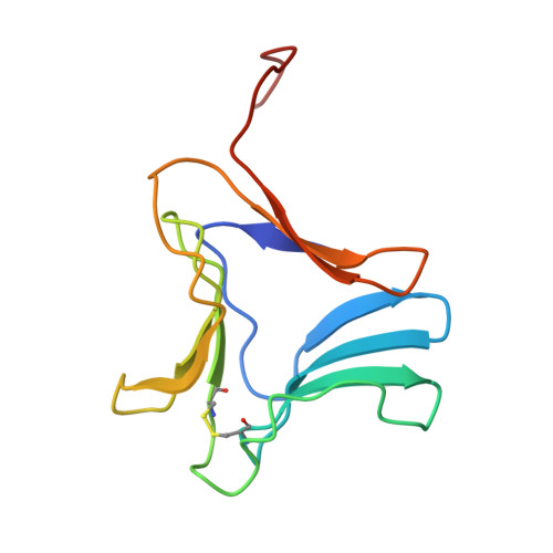
Galanthus nivalis agglutinin (GNA), a mannose-specific lectin from snowdrop bulbs, is a tetrameric member of the family of Amaryllidaceae lectins that exhibit antiviral activity towards HIV. Its subunits are composed of three pseudo-symmetrically related beta sheet domains, each with a conserved mannose-binding site. Crystal structures of monosaccharide and disaccharide complexes of GNA have revealed that all 12 binding sites of the tetramer are functional, and that the degree of occupancy is dependent on the availability of subsidiary interactions from neighboring subunits. The complex of GNA with a branched mannopentaose ((Manalpha1,6-(alpha1, 3-Man)Man-alpha1,6-(alpha1,3-Man)Man) described here simulates a more biologically relevant complex. Two unique mannopentaose binding modes co-exist in the tetragonal structure (1 subunit/asymmetric unit) of the complex. In one, the conserved monosaccharide-binding pocket in domain 1 (CRD 1) is utilized for cross-linkage of twofold related GNA dimers by the outer 3,6 tri-Man arm, which alternates between two orientations consistent with crystal symmetry. Inter-linked dimers assemble helically along the 41 crystal axis forming a pore-like structure. In the second binding mode, the complete 3,6 tri-Man arm binds to an extended binding region in domain 3 (CRD 3) with subsites for each terminal Man and the internal Man positioned in the conserved monosaccharide pocket. The two remaining mannose residues are not visible in either binding mode. This structure provides insights into possible mechanisms of the cross-linkage that is known to occur when lectins interact with specific multivalent cell surface receptors during events such as agglutination and mitogenic stimulation. By virtue of the large number of sites available for mannose binding, GNA has multiple possibilities of forming unique lattice structures. The two distinctly different binding modes observed in this study confirm that high affinity mannose binding occurs only at the two domain sites located near dimer interfaces.
- Department of Biochemistry, Virginia Commonwealth University, Richmond, Virginia 23298, USA. cswright@gems.vcu.edu
Organizational Affiliation: