1JOI | pdb_00001joi

STRUCTURE OF PSEUDOMONAS FLUORESCENS HOLO AZURIN


Experimental Data Snapshot

  • Method: X-RAY DIFFRACTION
  • Resolution: 2.05 Å
  • R-Value Free: 
    0.241 (Depositor), 0.220 (DCC) 
  • R-Value Work: 
    0.170 (Depositor), 0.157 (DCC) 
  • R-Value Observed: 
    0.170 (Depositor) 

Starting Model: experimental
View more details

wwPDB Validation 3D Report Full Report

Validation slider image for 1JOI

This is version 1.5 of the entry. See complete history

Literature

Crystallization and preliminary crystallographic studies of the crystals of the azurin Pseudomonas fluorescens.

Zhu, D.W.Dahms, T.Willis, K.Szabo, A.G.Lee, X.

(1994) Arch Biochem Biophys 308: 469-470

  • DOI: https://doi.org/10.1006/abbi.1994.1066
  • Primary Citation Related Structures: 
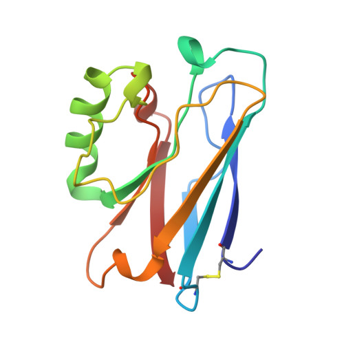
    1JOI

  • PubMed Abstract: 

    The Azurin Pseudomones fluorescens has been crystallized in the presence of ammonium sulfate and Tris buffer at pH 7.5. The crystals diffract to 2.05 A using a FAST system. The space group is P2(1)2(1)2(1) with a = 31.95, b = 43.78, and c = 78.81 A.


  • Organizational Affiliation
    • MRC Group in Molecular Endocrinology, CHUL Research Center, Ste-Foy, Québec, Canada.

Macromolecule Content 

  • Total Structure Weight: 13.71 kDa 
  • Atom Count: 1,066 
  • Modeled Residue Count: 128 
  • Deposited Residue Count: 128 
  • Unique protein chains: 1

Macromolecules

Find similar proteins by:|  3D Structure
Entity ID: 1
MoleculeChains  Sequence LengthOrganismDetailsImage
AZURIN128Pseudomonas fluorescens bv. AMutation(s): 0 
UniProt
Find proteins for P80546 (Pseudomonas fluorescens biotype A)
Explore P80546 
Go to UniProtKB:  P80546
Entity Groups
Sequence Clusters30% Identity50% Identity70% Identity90% Identity95% Identity100% Identity
UniProt GroupP80546
Sequence Annotations
Expand
Reference Sequence

Small Molecules

Ligands 1 Unique
IDChains Name / Formula / InChI Key2D Diagram3D Interactions
CU

Query on CU


Download:Ideal Coordinates CCD File
B [auth A]COPPER (II) ION
Cu
JPVYNHNXODAKFH-UHFFFAOYSA-N

Experimental Data & Validation

Experimental Data

  • Method: X-RAY DIFFRACTION
  • Resolution: 2.05 Å
  • R-Value Free:  0.241 (Depositor), 0.220 (DCC) 
  • R-Value Work:  0.170 (Depositor), 0.157 (DCC) 
  • R-Value Observed: 0.170 (Depositor) 
Space Group: P 21 21 21
Unit Cell:
Length ( Å )Angle ( ˚ )
a = 31.95α = 90
b = 43.78β = 90
c = 78.81γ = 90
Software Package:
Software NamePurpose
MADNESdata collection
CCP4data reduction
X-PLORmodel building
X-PLORrefinement
MADNESdata reduction
CCP4data scaling
X-PLORphasing

Structure Validation

View Full Validation Report


Entry History 

Deposition Data

Revision History  (Full details and data files)

  • Version 1.0: 1997-12-10
    Type: Initial release
  • Version 1.1: 2008-03-24
    Changes: Version format compliance
  • Version 1.2: 2011-07-13
    Changes: Version format compliance
  • Version 1.3: 2018-04-18
    Changes: Data collection, Other
  • Version 1.4: 2023-08-09
    Changes: Database references, Derived calculations, Refinement description
  • Version 1.5: 2024-11-06
    Changes: Data collection, Structure summary