1JO6 | pdb_00001jo6

Solution structure of the cytoplasmic N-terminus of the BK beta-subunit KCNMB2


Experimental Data Snapshot

  • Method: SOLUTION NMR
  • Conformers Calculated: 30 
  • Conformers Submitted: 24 
  • Selection Criteria: structures with the least restraint violations,target function 

wwPDB Validation 3D Report Full Report

Validation slider image for 1JO6

This is version 1.4 of the entry. See complete history

Literature

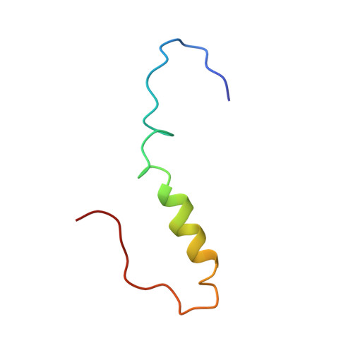
NMR structure of the "ball-and-chain" domain of KCNMB2, the beta 2-subunit of large conductance Ca2+- and voltage-activated potassium channels.

Bentrop, D.Beyermann, M.Wissmann, R.Fakler, B.

(2001) J Biological Chem 276: 42116-42121

  • DOI: https://doi.org/10.1074/jbc.M107118200
  • Primary Citation Related Structures: 
    1JO6

  • PubMed Abstract: 

    The auxiliary beta-subunit KCNMB2 (beta(2)) endows the non-inactivating large conductance Ca(2+)- and voltage-dependent potassium (BK) channel with fast inactivation. This process is mediated by the N terminus of KCNMB2 and closely resembles the "ball-and-chain"-type inactivation observed in voltage-gated potassium channels. Here we investigated the solution structure and function of the KCNMB2 N terminus (amino acids 1-45, BKbeta(2)N) using NMR spectroscopy and patch clamp recordings. BKbeta(2)N completely inactivated BK channels when applied to the cytoplasmic side; its interaction with the BK alpha-subunit is characterized by a particularly slow dissociation rate and an affinity in the upper nanomolar range. The BKbeta(2)N structure comprises two domains connected by a flexible linker: the pore-blocking "ball domain" (formed by residues 1-17) and the "chain domain" (between residues 20-45) linking it to the membrane segment of KCNMB2. The ball domain is made up of a flexible N terminus anchored at a well ordered loop-helix motif. The chain domain consists of a 4-turn helix with an unfolded linker at its C terminus. These structural properties explain the functional characteristics of BKbeta(2)N-mediated inactivation.


  • Organizational Affiliation
    • Department of Physiology II, University of Tübingen, Ob dem Himmelreich 7, 72074 Tübingen, Germany. detlef.bentrop@uni-tuebingen.de

Macromolecule Content 

  • Total Structure Weight: 5.38 kDa 
  • Atom Count: 377 
  • Modeled Residue Count: 45 
  • Deposited Residue Count: 45 
  • Unique protein chains: 1

Macromolecules

Find similar proteins by:|  3D Structure
Entity ID: 1
MoleculeChains  Sequence LengthOrganismDetailsImage
potassium large conductance calcium-activated channel, subfamily M, beta member 245N/AMutation(s): 0 
UniProt & NIH Common Fund Data Resources
Find proteins for Q9Y691 (Homo sapiens)
Explore Q9Y691 
Go to UniProtKB:  Q9Y691
PHAROS:  Q9Y691
GTEx:  ENSG00000197584 
Entity Groups
Sequence Clusters30% Identity50% Identity70% Identity90% Identity95% Identity100% Identity
UniProt GroupQ9Y691
Sequence Annotations
Expand
Reference Sequence

Experimental Data & Validation

Experimental Data

  • Method: SOLUTION NMR
  • Conformers Calculated: 30 
  • Conformers Submitted: 24 
  • Selection Criteria: structures with the least restraint violations,target function 

Structure Validation

View Full Validation Report


Entry History 

Deposition Data

Revision History  (Full details and data files)

  • Version 1.0: 2001-11-16
    Type: Initial release
  • Version 1.1: 2008-04-27
    Changes: Version format compliance
  • Version 1.2: 2011-07-13
    Changes: Version format compliance
  • Version 1.3: 2022-02-23
    Changes: Data collection, Database references, Derived calculations
  • Version 1.4: 2024-05-22
    Changes: Data collection