1JND | pdb_00001jnd

Crystal structure of imaginal disc growth factor-2


Experimental Data Snapshot

  • Method: X-RAY DIFFRACTION
  • Resolution: 1.30 Å
  • R-Value Free: 
    0.202 (Depositor), 0.193 (DCC) 
  • R-Value Work: 
    0.176 (Depositor), 0.167 (DCC) 
  • R-Value Observed: 
    0.176 (Depositor) 

Starting Model: experimental
View more details

wwPDB Validation 3D Report Full Report

Validation slider image for 1JND

This is version 2.2 of the entry. See complete history

Literature

Crystal structure of imaginal disc growth factor-2. A member of a new family of growth-promoting glycoproteins from Drosophila melanogaster.

Varela, P.F.Llera, A.S.Mariuzza, R.A.Tormo, J.

(2002) J Biological Chem 277: 13229-13236

  • DOI: https://doi.org/10.1074/jbc.M110502200
  • Primary Citation Related Structures: 
    1JND, 1JNE

  • PubMed Abstract: 

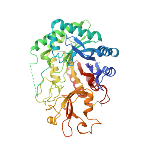
    Imaginal disc growth factor-2 (IDGF-2) is a member of a recently described family of Drosophila melanogaster-soluble polypeptide growth factors that promote cell proliferation in imaginal discs. Although their precise mode of action has not been established, IDGFs cooperate with insulin in stimulating the growth of imaginal disc cells. We report the crystal structure of IDGF-2 at 1.3-A resolution. The structure shows the classical (betaalpha)(8) barrel-fold of family 18 glycosyl hydrolases, with an insertion of an alpha + beta domain similar to that of Serratia marcescens chitinases A and B. However, amino acid substitutions in the consensus catalytic sequence of chitinases give IDGF-2 a less negatively charged environment in its putative ligand-binding site and preclude the nucleophilic attack mechanism of chitin hydrolysis. Particularly important is the replacement of Glu by Gln at position 132, which has been shown to abolish enzymatic activity in chitinases. Nevertheless, a modest conservation of residues that participate in oligosaccharide recognition suggests that IDGF-2 could bind carbohydrates, assuming several conformational changes to open the partially occluded binding site. Thus, IDGFs may have evolved from chitinases to acquire new functions as growth factors, interacting with cell surface glycoproteins implicated in growth-promoting processes, such as the Drosophila insulin receptor.


  • Organizational Affiliation
    • Center for Advanced Research in Biotechnology, W. M. Keck Laboratory of Structural Biology, University of Maryland Biotechnology Institute, Rockville, MD 20850, USA.

Macromolecule Content 

  • Total Structure Weight: 47.7 kDa 
  • Atom Count: 3,916 
  • Modeled Residue Count: 400 
  • Deposited Residue Count: 420 
  • Unique protein chains: 1

Macromolecules

Find similar proteins by:|  3D Structure
Entity ID: 1
MoleculeChains  Sequence LengthOrganismDetailsImage
Imaginal disc growth factor-2420Drosophila melanogasterMutation(s): 0 
UniProt
Find proteins for Q9V3D4 (Drosophila melanogaster)
Explore Q9V3D4 
Go to UniProtKB:  Q9V3D4
Entity Groups
Sequence Clusters30% Identity50% Identity70% Identity90% Identity95% Identity100% Identity
UniProt GroupQ9V3D4
Glycosylation
Glycosylation Sites: 1Go to GlyGen: Q9V3D4-1
Sequence Annotations
Expand
Reference Sequence

Oligosaccharides

Help  
Entity ID: 2
MoleculeChains Length2D Diagram GlycosylationD Interactions
alpha-D-mannopyranose-(1-3)-beta-D-mannopyranose-(1-4)-2-acetamido-2-deoxy-beta-D-glucopyranose-(1-4)-2-acetamido-2-deoxy-beta-D-glucopyranose
B
4N-Glycosylation
Glycosylation Resources
GlyTouCan: G81315DD
GlyCosmos: G81315DD
GlyGen: G81315DD

Experimental Data & Validation

Experimental Data

  • Method: X-RAY DIFFRACTION
  • Resolution: 1.30 Å
  • R-Value Free:  0.202 (Depositor), 0.193 (DCC) 
  • R-Value Work:  0.176 (Depositor), 0.167 (DCC) 
  • R-Value Observed: 0.176 (Depositor) 
Space Group: P 32 2 1
Unit Cell:
Length ( Å )Angle ( ˚ )
a = 106.345α = 90
b = 106.345β = 90
c = 89.987γ = 120
Software Package:
Software NamePurpose
DENZOdata reduction
SCALEPACKdata scaling
AMoREphasing
SHELXL-97refinement

Structure Validation

View Full Validation Report


Entry History 

Deposition Data

Revision History  (Full details and data files)

  • Version 1.0: 2002-05-01
    Type: Initial release
  • Version 1.1: 2007-10-21
    Changes: Version format compliance
  • Version 1.2: 2011-07-13
    Changes: Non-polymer description, Version format compliance
  • Version 2.0: 2020-07-29
    Type: Remediation
    Reason: Carbohydrate remediation
    Changes: Advisory, Atomic model, Data collection, Database references, Derived calculations, Structure summary
  • Version 2.1: 2023-08-16
    Changes: Data collection, Database references, Refinement description, Structure summary
  • Version 2.2: 2024-11-13
    Changes: Structure summary