1JN0 | pdb_00001jn0

Crystal structure of the non-regulatory A4 isoform of spinach chloroplast glyceraldehyde-3-phosphate dehydrogenase complexed with NADP


Experimental Data Snapshot

  • Method: X-RAY DIFFRACTION
  • Resolution: 3.00 Å
  • R-Value Free: 
    0.254 (Depositor) 
  • R-Value Work: 
    0.213 (Depositor) 
  • R-Value Observed: 
    0.213 (Depositor) 

Starting Model: experimental
View more details

wwPDB Validation 3D Report Full Report

Validation slider image for 1JN0

This is version 1.5 of the entry. See complete history

Literature

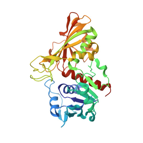
Crystal structure of the non-regulatory A(4 )isoform of spinach chloroplast glyceraldehyde-3-phosphate dehydrogenase complexed with NADP.

Fermani, S.Ripamonti, A.Sabatino, P.Zanotti, G.Scagliarini, S.Sparla, F.Trost, P.Pupillo, P.

(2001) J Mol Biology 314: 527-542

  • DOI: https://doi.org/10.1006/jmbi.2001.5172
  • Primary Citation Related Structures: 
    1JN0

  • PubMed Abstract: 

    Here, we report the first crystal structure of a photosynthetic glyceraldehyde-3-phosphate dehydrogenase (GAPDH) complexed with NADP. The enzyme, purified from spinach chloroplasts, is constituted of a single type of subunit (A) arranged in homotetramers. It shows non-regulated NADP-dependent and NAD-dependent activities, with a preference for NADP. The structure has been solved to 3.0 A resolution by molecular replacement. The crystals belong to space group C222 with three monomers in the asymmetric unit. One of the three monomers generates a tetramer using the space group 222 point symmetry and a very similar tetramer is generated by the other two monomers, related by a non-crystallographic symmetry, using a crystallographic 2-fold axis. The protein reveals a large structural homology with known GAPDHs both in the cofactor-binding domain and in regions of the catalytic domain. Like all other GAPDHs investigated so far, the A(4)-GAPDH belongs to the Rossmann fold family of dehydrogenases. However, unlike most dehydrogenases of this family, the adenosine 2'-phosphate group of NADP does not form a salt-bridge with any positively charged residue in its surroundings, being instead set in place by hydrogen bonds with a threonine residue belonging to the Rossmann fold and a serine residue located in the S-loop of a symmetry-related monomer. While increasing our knowledge of an important photosynthetic enzyme, these results contribute to a general understanding of NADP versus NAD recognition in pyridine nucleotide-dependent enzymes. Although the overall structure of A(4)-GAPDH is similar to that of the cytosolic GAPDH from bacteria and eukaryotes, the chloroplast tetramer is peculiar, in that it can actually be considered a dimer of dimers, since monomers are bound in pairs by a disulphide bridge formed across Cys200 residues. This bridge is not found in other cytosolic or chloroplast GAPDHs from animals, bacteria, or plants other than spinach.


  • Organizational Affiliation
    • Dipartimento di Chimica "G. Ciamician", Università di Bologna, via Selmi 2, Bologna, 40126, Italia. fermani@ciam.unibo.it

Macromolecule Content 

  • Total Structure Weight: 110.85 kDa 
  • Atom Count: 7,949 
  • Modeled Residue Count: 1,002 
  • Deposited Residue Count: 1,005 
  • Unique protein chains: 1

Macromolecules

Find similar proteins by:|  3D Structure
Entity ID: 1
MoleculeChains  Sequence LengthOrganismDetailsImage
GLYCERALDEHYDE-3-PHOSPHATE DEHYDROGENASE AA [auth O],
B [auth A],
C [auth B]
335Spinacia oleraceaMutation(s): 0 
EC: 1.2.1.13
UniProt
Find proteins for P19866 (Spinacia oleracea)
Explore P19866 
Go to UniProtKB:  P19866
Entity Groups
Sequence Clusters30% Identity50% Identity70% Identity90% Identity95% Identity100% Identity
UniProt GroupP19866
Sequence Annotations
Expand
Reference Sequence

Small Molecules

Ligands 2 Unique
IDChains Name / Formula / InChI Key2D Diagram3D Interactions
NDP

Query on NDP


Download:Ideal Coordinates CCD File
F [auth O],
I [auth A],
L [auth B]
NADPH DIHYDRO-NICOTINAMIDE-ADENINE-DINUCLEOTIDE PHOSPHATE
C21 H30 N7 O17 P3
ACFIXJIJDZMPPO-NNYOXOHSSA-N
SO4

Query on SO4


Download:Ideal Coordinates CCD File
D [auth O]
E [auth O]
G [auth A]
H [auth A]
J [auth B]
D [auth O],
E [auth O],
G [auth A],
H [auth A],
J [auth B],
K [auth B]
SULFATE ION
O4 S
QAOWNCQODCNURD-UHFFFAOYSA-L

Experimental Data & Validation

Experimental Data

  • Method: X-RAY DIFFRACTION
  • Resolution: 3.00 Å
  • R-Value Free:  0.254 (Depositor) 
  • R-Value Work:  0.213 (Depositor) 
  • R-Value Observed: 0.213 (Depositor) 
Space Group: C 2 2 2
Unit Cell:
Length ( Å )Angle ( ˚ )
a = 146.249α = 90
b = 182.184β = 90
c = 106.026γ = 90
Software Package:
Software NamePurpose
DENZOdata reduction
SCALEPACKdata scaling
AMoREphasing
CNSrefinement

Structure Validation

View Full Validation Report


Entry History 

Deposition Data

Revision History  (Full details and data files)

  • Version 1.0: 2001-11-30
    Type: Initial release
  • Version 1.1: 2008-04-27
    Changes: Version format compliance
  • Version 1.2: 2011-07-13
    Changes: Derived calculations, Version format compliance
  • Version 1.3: 2014-11-12
    Changes: Structure summary
  • Version 1.4: 2023-08-16
    Changes: Data collection, Database references, Derived calculations, Refinement description
  • Version 1.5: 2024-10-09
    Changes: Structure summary