1JM1 | pdb_00001jm1

Crystal structure of the soluble domain of the Rieske protein II (soxF) from Sulfolobus acidocaldarius


Experimental Data Snapshot

  • Method: X-RAY DIFFRACTION
  • Resolution: 1.11 Å
  • R-Value Free: 
    0.125 (Depositor) 
  • R-Value Work: 
    0.106 (Depositor), 0.095 (DCC) 
  • R-Value Observed: 
    0.106 (Depositor) 

wwPDB Validation 3D Report Full Report

Validation slider image for 1JM1

This is version 1.3 of the entry. See complete history

Literature

The structure of the soluble domain of an archaeal Rieske iron-sulfur protein at 1.1 A resolution.

Bonisch, H.Schmidt, C.L.Schafer, G.Ladenstein, R.

(2002) J Mol Biology 319: 791-805

  • DOI: https://doi.org/10.1016/S0022-2836(02)00323-6
  • Primary Citation Related Structures: 
    1JM1

  • PubMed Abstract: 

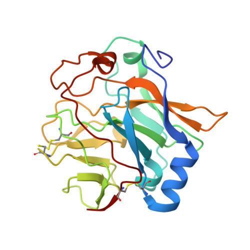
    The first crystal structure of an archaeal Rieske iron-sulfur protein, the soluble domain of Rieske iron-sulfur protein II (soxF) from the hyperthermo-acidophile Sulfolobus acidocaldarius, has been solved by multiple wavelength anomalous dispersion (MAD) and has been refined to 1.1 A resolution. SoxF is a subunit of the terminal oxidase supercomplex SoxM in the plasma membrane of S. acidocaldarius that combines features of a cytochrome bc(1) complex and a cytochrome c oxidase. The [2Fe-2S] cluster of soxF is most likely the primary electron acceptor during the oxidation of caldariella quinone by the cytochrome a(587)/Rieske subcomplex. The geometry of the [2Fe-2S] cluster and the structure of the cluster-binding site are almost identical in soxF and the Rieske proteins from eucaryal cytochrome bc(1) and b(6)f complexes, suggesting a strict conservation of the catalytic mechanism. The main domain of soxF and part of the cluster-binding domain, though structurally related, show a significantly divergent structure with respect to topology, non-covalent interactions and surface charges. The divergent structure of soxF reflects a different topology of the soxM complex compared to eucaryal bc complexes and the adaptation of the protein to the extreme ambient conditions on the outer membrane surface of a hyperthermo-acidophilic organism.


  • Organizational Affiliation
    • Department of Biosciences at NOVUM, Center for Structural Biochemistry, Karolinska Institutet, Hälsovägen 7-9, S-14157 Huddinge, Sweden.

Macromolecule Content 

  • Total Structure Weight: 21.9 kDa 
  • Atom Count: 1,893 
  • Modeled Residue Count: 202 
  • Deposited Residue Count: 204 
  • Unique protein chains: 1

Macromolecules

Find similar proteins by:|  3D Structure
Entity ID: 1
MoleculeChains  Sequence LengthOrganismDetailsImage
Rieske iron-sulfur protein soxF204Sulfolobus acidocaldariusMutation(s): 0 
Gene Names: SOXF
UniProt
Find proteins for Q53766 (Sulfolobus acidocaldarius)
Explore Q53766 
Go to UniProtKB:  Q53766
Entity Groups
Sequence Clusters30% Identity50% Identity70% Identity90% Identity95% Identity100% Identity
UniProt GroupQ53766
Sequence Annotations
Expand
Reference Sequence

Experimental Data & Validation

Experimental Data

  • Method: X-RAY DIFFRACTION
  • Resolution: 1.11 Å
  • R-Value Free:  0.125 (Depositor) 
  • R-Value Work:  0.106 (Depositor), 0.095 (DCC) 
  • R-Value Observed: 0.106 (Depositor) 
Space Group: P 61
Unit Cell:
Length ( Å )Angle ( ˚ )
a = 80.252α = 90
b = 80.252β = 90
c = 75.624γ = 120
Software Package:
Software NamePurpose
DENZOdata reduction
SCALEPACKdata scaling
MLPHAREphasing
SHELXL-97refinement

Structure Validation

View Full Validation Report


Entry History 

Deposition Data

Revision History  (Full details and data files)

  • Version 1.0: 2002-07-17
    Type: Initial release
  • Version 1.1: 2008-04-27
    Changes: Version format compliance
  • Version 1.2: 2011-07-13
    Changes: Version format compliance
  • Version 1.3: 2024-11-13
    Changes: Data collection, Database references, Derived calculations, Structure summary