1JKI

myo-Inositol-1-phosphate Synthase Complexed with an Inhibitor, 2-deoxy-glucitol-6-phosphate


Experimental Data Snapshot

  • Method: X-RAY DIFFRACTION
  • Resolution: 2.20 Å
  • R-Value Free: 0.280 
  • R-Value Work: 0.208 
  • R-Value Observed: 0.214 

wwPDB Validation   3D Report Full Report


This is version 1.3 of the entry. See complete history


Literature

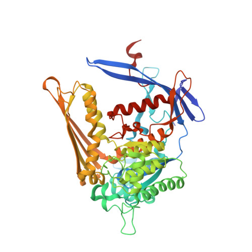
The crystal structure and mechanism of 1-L-myo-inositol- 1-phosphate synthase

Stein, A.J.Geiger, J.H.

(2002) J Biol Chem 277: 9484-9491

  • DOI: https://doi.org/10.1074/jbc.M109371200
  • Primary Citation of Related Structures:  
    1JKF, 1JKI

  • PubMed Abstract: 

    1-l-myo-Inositol-1-phosphate synthase catalyzes the conversion of d-glucose 6-phosphate to 1-l-myo-inositol-1-phosphate (MIP), the first and rate-limiting step in the biosynthesis of all inositol-containing compounds. It involves an oxidation, intramolecular aldol cyclization, and reduction. We have determined the first crystal structure of MIP synthase. We present structures of both the NAD-bound enzyme and the enzyme bound to an inhibitor, 2-deoxy-glucitol-6-phosphate. While 58 amino acids are disordered in the unbound form of the enzyme in the vicinity of the active site, the inhibitor nucleates the folding of this domain in a striking example of induced fit, serving to completely encapsulate it within the enzyme. Three helices and a long beta-strand are formed in this process. We postulate a mechanism for the conversion based on the structure of the inhibitor-bound complex.


  • Organizational Affiliation

    Department of Chemistry, Michigan State University, East Lansing, Michigan 48824, USA.


Macromolecules
Find similar proteins by:  (by identity cutoff)  |  3D Structure
Entity ID: 1
MoleculeChains Sequence LengthOrganismDetailsImage
myo-inositol-1-phosphate synthase
A, B
533Saccharomyces cerevisiaeMutation(s): 0 
EC: 5.5.1.4
UniProt
Find proteins for P11986 (Saccharomyces cerevisiae (strain ATCC 204508 / S288c))
Explore P11986 
Go to UniProtKB:  P11986
Entity Groups  
Sequence Clusters30% Identity50% Identity70% Identity90% Identity95% Identity100% Identity
UniProt GroupP11986
Sequence Annotations
Expand
  • Reference Sequence
Small Molecules
Binding Affinity Annotations 
IDSourceBinding Affinity
DG6 Binding MOAD:  1JKI Ki: 6000 (nM) from 1 assay(s)
Experimental Data & Validation

Experimental Data

  • Method: X-RAY DIFFRACTION
  • Resolution: 2.20 Å
  • R-Value Free: 0.280 
  • R-Value Work: 0.208 
  • R-Value Observed: 0.214 
  • Space Group: C 1 2 1
Unit Cell:
Length ( Å )Angle ( ˚ )
a = 152.728α = 90
b = 98.309β = 126.093
c = 121.855γ = 90
Software Package:
Software NamePurpose
CNSrefinement
DENZOdata reduction
SCALEPACKdata scaling
CNSphasing

Structure Validation

View Full Validation Report


Entry History 

Deposition Data

Revision History  (Full details and data files)

  • Version 1.0: 2002-04-10
    Type: Initial release
  • Version 1.1: 2008-04-27
    Changes: Version format compliance
  • Version 1.2: 2011-07-13
    Changes: Derived calculations, Version format compliance
  • Version 1.3: 2023-08-16
    Changes: Data collection, Database references, Derived calculations, Refinement description