1J88 | pdb_00001j88

HUMAN HIGH AFFINITY FC RECEPTOR FC(EPSILON)RI(ALPHA), TETRAGONAL CRYSTAL FORM 1


Experimental Data Snapshot

  • Method: X-RAY DIFFRACTION
  • Resolution: 3.20 Å
  • R-Value Free: 
    0.310 (Depositor) 
  • R-Value Work: 
    0.262 (Depositor) 
  • R-Value Observed: 
    0.262 (Depositor) 

Starting Model: experimental
View more details

wwPDB Validation 3D Report Full Report

Validation slider image for 1J88

This is version 2.2 of the entry. See complete history

Literature

The analysis of the human high affinity IgE receptor Fc epsilon Ri alpha from multiple crystal forms.

Garman, S.C.Sechi, S.Kinet, J.P.Jardetzky, T.S.

(2001) J Mol Biology 311: 1049-1062

  • DOI: https://doi.org/10.1006/jmbi.2001.4929
  • Primary Citation Related Structures: 
    1J86, 1J87, 1J88, 1J89

  • PubMed Abstract: 

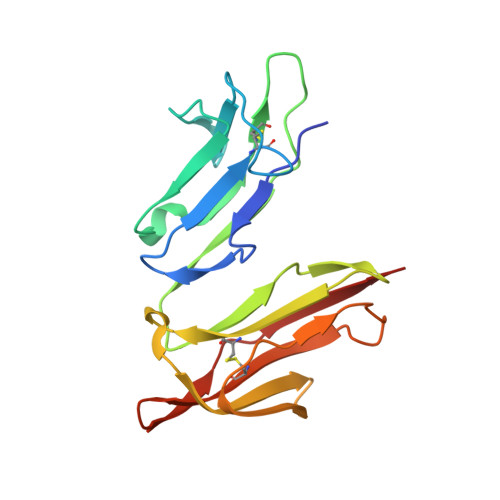
    We have solved the structure of the human high affinity IgE receptor, Fc epsilon RI alpha, in six different crystal forms, showing the structure in 15 different chemical environments. This database of structures shows no change in the overall shape of the molecule, as the angle between domains 1 and 2 (D1 and D2) varies little across the ensemble. However, the receptor has local conformational variability in the C' strand of D2 and in the BC loop of D1. In every crystal form, a residue inserts between tryptophan residues 87 and 110, mimicking the position of a proline from the IgE ligand. The different crystal forms reveal a distribution of carbohydrates lining the front and back surfaces of the structure. An analysis of crystal contacts in the different forms indicates regions where the molecule interacts with other proteins, and reveals a potential new binding site distal to the IgE binding site. The results of this study point to new directions for the design of molecules to inhibit the interaction of Fc epsilon RI alpha with its natural ligand and thus to prevent a primary step in the allergic response.


  • Organizational Affiliation
    • Laboratory of Immunogenetics, National Institute of Allergy and Infectious Diseases, National Institutes of Health, Twinbrook II, 12441 Parklawn Drive, Rockville, MD 20852, USA. garman@alpha.niaid.nih.gov

Macromolecule Content 

  • Total Structure Weight: 110.49 kDa 
  • Atom Count: 7,606 
  • Modeled Residue Count: 840 
  • Deposited Residue Count: 860 
  • Unique protein chains: 1

Macromolecules

Find similar proteins by:|  3D Structure
Entity ID: 1
MoleculeChains  Sequence LengthOrganismDetailsImage
HIGH AFFINITY IMMUNOGLOBULIN EPSILON RECEPTOR ALPHA-SUBUNIT
A, B, C, D, E
172Homo sapiensMutation(s): 0 
UniProt & NIH Common Fund Data Resources
Find proteins for P12319 (Homo sapiens)
Explore P12319 
Go to UniProtKB:  P12319
PHAROS:  P12319
GTEx:  ENSG00000179639 
Entity Groups
Sequence Clusters30% Identity50% Identity70% Identity90% Identity95% Identity100% Identity
UniProt GroupP12319
Glycosylation
Glycosylation Sites: 6Go to GlyGen: P12319-1
Sequence Annotations
Expand
Reference Sequence

Oligosaccharides

Help  
Entity ID: 2
MoleculeChains Length2D Diagram GlycosylationD Interactions
beta-D-mannopyranose-(1-4)-2-acetamido-2-deoxy-beta-D-glucopyranose-(1-4)-2-acetamido-2-deoxy-beta-D-glucopyranose
F, J, M, O, P
3N-Glycosylation
Glycosylation Resources
GlyTouCan: G15407YE
GlyCosmos: G15407YE
GlyGen: G15407YE
Entity ID: 3
MoleculeChains Length2D Diagram GlycosylationD Interactions
alpha-D-mannopyranose-(1-3)-alpha-D-mannopyranose-(1-4)-2-acetamido-2-deoxy-beta-D-glucopyranose-(1-4)-2-acetamido-2-deoxy-beta-D-glucopyranose
G
4N-Glycosylation
Glycosylation Resources
GlyTouCan: G48068RF
GlyCosmos: G48068RF
GlyGen: G48068RF
Entity ID: 4
MoleculeChains Length2D Diagram GlycosylationD Interactions
2-acetamido-2-deoxy-beta-D-glucopyranose-(1-4)-2-acetamido-2-deoxy-beta-D-glucopyranose
H, I, K, L, N
H, I, K, L, N, Q
2N-Glycosylation
Glycosylation Resources
GlyTouCan: G42666HT
GlyCosmos: G42666HT
GlyGen: G42666HT
Entity ID: 5
MoleculeChains Length2D Diagram GlycosylationD Interactions
alpha-D-mannopyranose-(1-6)-beta-D-mannopyranose-(1-4)-2-acetamido-2-deoxy-beta-D-glucopyranose-(1-4)-2-acetamido-2-deoxy-beta-D-glucopyranose
R
4N-Glycosylation
Glycosylation Resources
GlyTouCan: G22573RC
GlyCosmos: G22573RC
GlyGen: G22573RC

Small Molecules

Ligands 1 Unique
IDChains Name / Formula / InChI Key2D Diagram3D Interactions
NAG

Query on NAG


Download:Ideal Coordinates CCD File
AA [auth C]
BA [auth C]
CA [auth D]
DA [auth D]
EA [auth D]
AA [auth C],
BA [auth C],
CA [auth D],
DA [auth D],
EA [auth D],
FA [auth E],
GA [auth E],
HA [auth E],
IA [auth E],
S [auth A],
T [auth A],
U [auth A],
V [auth B],
W [auth B],
X [auth B],
Y [auth B],
Z [auth C]
2-acetamido-2-deoxy-beta-D-glucopyranose
C8 H15 N O6
OVRNDRQMDRJTHS-FMDGEEDCSA-N

Experimental Data & Validation

Experimental Data

  • Method: X-RAY DIFFRACTION
  • Resolution: 3.20 Å
  • R-Value Free:  0.310 (Depositor) 
  • R-Value Work:  0.262 (Depositor) 
  • R-Value Observed: 0.262 (Depositor) 
Space Group: P 43
Unit Cell:
Length ( Å )Angle ( ˚ )
a = 145.08α = 90
b = 145.08β = 90
c = 62.74γ = 90
Software Package:
Software NamePurpose
DENZOdata reduction
SCALEPACKdata scaling
AMoREphasing
CNSrefinement

Structure Validation

View Full Validation Report


Entry History 

Deposition Data

Revision History  (Full details and data files)

  • Version 1.0: 2001-08-29
    Type: Initial release
  • Version 1.1: 2008-04-27
    Changes: Version format compliance
  • Version 1.2: 2011-07-13
    Changes: Non-polymer description, Version format compliance
  • Version 2.0: 2020-07-29
    Type: Remediation
    Reason: Carbohydrate remediation
    Changes: Atomic model, Data collection, Derived calculations, Structure summary
  • Version 2.1: 2023-08-16
    Changes: Data collection, Database references, Refinement description, Structure summary
  • Version 2.2: 2024-11-13
    Changes: Structure summary