1J4W | pdb_00001j4w

COMPLEX OF THE KH3 and KH4 DOMAINS OF FBP WITH A SINGLE_STRANDED 29mer DNA OLIGONUCLEOTIDE FROM THE FUSE ELEMENT OF THE C-MYC ONCOGENE


Experimental Data Snapshot

  • Method: SOLUTION NMR
  • Conformers Calculated: 80 
  • Conformers Submitted: 
  • Selection Criteria: REGULARIZED MEAN STRUCTURE 

wwPDB Validation 3D Report Full Report

Validation slider image for 1J4W

This is version 1.4 of the entry. See complete history

Literature

Structure and dynamics of KH domains from FBP bound to single-stranded DNA.

Braddock, D.T.Louis, J.M.Baber, J.L.Levens, D.Clore, G.M.

(2002) Nature 415: 1051-1056

  • DOI: https://doi.org/10.1038/4151051a
  • Primary Citation Related Structures: 
    1J4W

  • PubMed Abstract: 

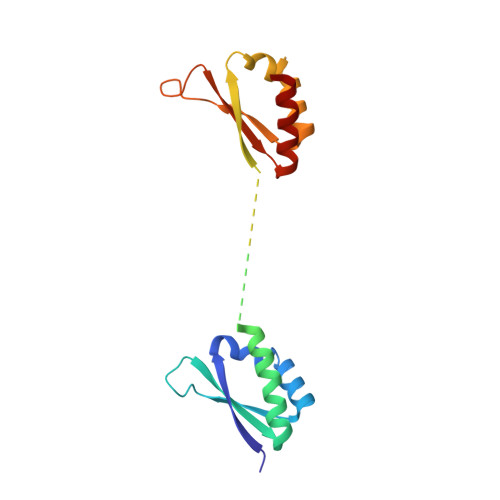
    Gene regulation can be tightly controlled by recognition of DNA deformations that are induced by stress generated during transcription. The KH domains of the FUSE-binding protein (FBP), a regulator of c-myc expression, bind in vivo and in vitro to the single-stranded far-upstream element (FUSE), 1,500 base pairs upstream from the c-myc promoter. FBP bound to FUSE acts through TFIIH at the promoter. Here we report the solution structure of a complex between the KH3 and KH4 domains of FBP and a 29-base single-stranded DNA from FUSE. The KH domains recognize two sites, 9-10 bases in length, separated by 5 bases, with KH4 bound to the 5' site and KH3 to the 3' site. The central portion of each site comprises a tetrad of sequence 5'd-ATTC for KH4 and 5'd-TTTT for KH3. Dynamics measurements show that the two KH domains bind as articulated modules to single-stranded DNA, providing a flexible framework with which to recognize transient, moving targets.


  • Organizational Affiliation
    • Laboratory of Chemical Physics, National Institute of Diabetes and Digestive and Kidney Diseases, National Institutes of Health, Bethesda, Maryland 20892, USA.

Macromolecule Content 

  • Total Structure Weight: 27.52 kDa 
  • Atom Count: 1,413 
  • Modeled Residue Count: 160 
  • Deposited Residue Count: 203 
  • Unique protein chains: 1
  • Unique nucleic acid chains: 1

Macromolecules


Find similar proteins by:|  3D Structure
Entity ID: 2
MoleculeChains  Sequence LengthOrganismDetailsImage
FUSE binding proteinB [auth A]174Homo sapiensMutation(s): 1 
UniProt & NIH Common Fund Data Resources
Find proteins for Q96AE4 (Homo sapiens)
Explore Q96AE4 
Go to UniProtKB:  Q96AE4
PHAROS:  Q96AE4
GTEx:  ENSG00000162613 
Entity Groups
Sequence Clusters30% Identity50% Identity70% Identity90% Identity95% Identity100% Identity
UniProt GroupQ96AE4
Sequence Annotations
Expand
Reference Sequence
Find similar nucleic acids by:  (by identity cutoff) 
Entity ID: 1
MoleculeChains LengthOrganismImage
DNA (5'-D(*GP*TP*A*TP*AP*TP*TP*CP*CP*CP*TP*CP*GP*GP*G*AP*TP*TP*TP*TP*TP*TP*AP*TP*TP*TP*TP*GP*T)-3')A [auth B]29N/A
Sequence Annotations
Expand
Reference Sequence

Experimental Data & Validation

Experimental Data

  • Method: SOLUTION NMR
  • Conformers Calculated: 80 
  • Conformers Submitted: 
  • Selection Criteria: REGULARIZED MEAN STRUCTURE 

Structure Validation

View Full Validation Report


Entry History 

Deposition Data

Revision History  (Full details and data files)

  • Version 1.0: 2002-03-06
    Type: Initial release
  • Version 1.1: 2008-04-26
    Changes: Version format compliance
  • Version 1.2: 2011-07-13
    Changes: Version format compliance
  • Version 1.3: 2021-10-27
    Changes: Data collection, Database references, Derived calculations
  • Version 1.4: 2023-12-27
    Changes: Data collection