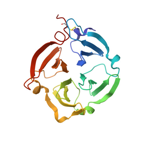
1ITV

Dimeric form of the haemopexin domain of MMP9


Experimental Data Snapshot

  • Method: X-RAY DIFFRACTION
  • Resolution: 1.95 Å
  • R-Value Free: 0.275 
  • R-Value Work: 0.229 
  • R-Value Observed: 0.232 

wwPDB Validation   3D Report Full Report


This is version 1.3 of the entry. See complete history


Literature

Structural basis of the adaptive molecular recognition by MMP9.

Cha, H.Kopetzki, E.Huber, R.Lanzendorfer, M.Brandstetter, H.

(2002) J Mol Biol 320: 1065-1079

  • DOI: https://doi.org/10.1016/s0022-2836(02)00558-2
  • Primary Citation of Related Structures:  
    1ITV

  • PubMed Abstract: 

    Matrix metalloproteinase (MMPs) are critical for the degradation of extracellular matrix components and, therefore, need to be regulated tightly. Almost all MMPs share a homologous C-terminal haemopexin-like domain (PEX). Besides its role in macromolecular substrate processing, the PEX domains appear to play a major role in regulating MMP activation, localisation and inhibition. One intriguing property of MMP9 is its competence to bind different proteins, involved in these regulatory processes, with high affinity at an overlapping recognition site on its PEX domain. With the crystal structure of the PEX9 dimer, we present the first example of how PEX domains accomplish these diverse roles. Blade IV of PEX9 mediates the non-covalent and predominantly hydrophobic dimerisation contact. Large shifts of blade III and, in particular, blade IV, accompany the dimerisation, resulting in a remarkably asymmetric homodimeric structure. The asymmetry provides a novel mechanism of adaptive protein recognition, where different proteins (PEX9, PEX1, and TIMP1) can bind with high affinity to PEX9 at an overlapping site. Finally, the structure illustrates how the dimerisation generates new properties on both a physico-chemical and functional level.


  • Organizational Affiliation

    Max-Planck-Institut für Biochemie, Abteilung Strukturforschung, D-82152, Martinsried, Germany.


Macromolecules
Find similar proteins by:  (by identity cutoff)  |  3D Structure
Entity ID: 1
MoleculeChains Sequence LengthOrganismDetailsImage
MMP9
A, B
195Homo sapiensMutation(s): 0 
EC: 3.4.24.35
UniProt & NIH Common Fund Data Resources
Find proteins for P14780 (Homo sapiens)
Explore P14780 
Go to UniProtKB:  P14780
PHAROS:  P14780
GTEx:  ENSG00000100985 
Entity Groups  
Sequence Clusters30% Identity50% Identity70% Identity90% Identity95% Identity100% Identity
UniProt GroupP14780
Sequence Annotations
Expand
  • Reference Sequence
Experimental Data & Validation

Experimental Data

  • Method: X-RAY DIFFRACTION
  • Resolution: 1.95 Å
  • R-Value Free: 0.275 
  • R-Value Work: 0.229 
  • R-Value Observed: 0.232 
  • Space Group: P 43
Unit Cell:
Length ( Å )Angle ( ˚ )
a = 127.72α = 90
b = 127.72β = 90
c = 31.38γ = 90
Software Package:
Software NamePurpose
DENZOdata reduction
SCALEPACKdata scaling
MLPHAREphasing
MAINmodel building
SOLOMONphasing
CNSrefinement
MAINphasing

Structure Validation

View Full Validation Report


Entry History 

Deposition Data

Revision History  (Full details and data files)

  • Version 1.0: 2002-09-04
    Type: Initial release
  • Version 1.1: 2008-04-27
    Changes: Version format compliance
  • Version 1.2: 2011-07-13
    Changes: Version format compliance
  • Version 1.3: 2023-12-27
    Changes: Data collection, Database references, Derived calculations