1IT6

CRYSTAL STRUCTURE OF THE COMPLEX BETWEEN CALYCULIN A AND THE CATALYTIC SUBUNIT OF PROTEIN PHOSPHATASE 1


Experimental Data Snapshot

  • Method: X-RAY DIFFRACTION
  • Resolution: 2.00 Å
  • R-Value Free: 0.218 
  • R-Value Work: 0.182 

wwPDB Validation   3D Report Full Report


This is version 1.3 of the entry. See complete history


Literature

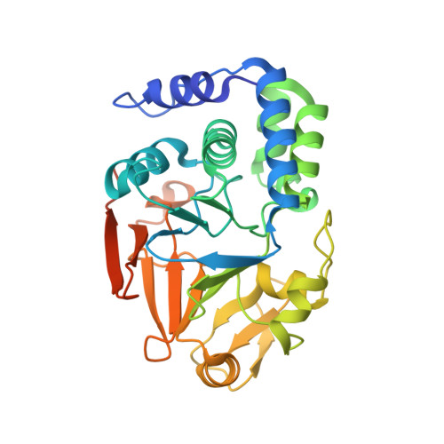
Crystal structure of the complex between calyculin A and the catalytic subunit of protein phosphatase 1.

Kita, A.Matsunaga, S.Takai, A.Kataiwa, H.Wakimoto, T.Fusetani, N.Isobe, M.Miki, K.

(2002) Structure 10: 715-724

  • DOI: https://doi.org/10.1016/s0969-2126(02)00764-5
  • Primary Citation of Related Structures:  
    1IT6

  • PubMed Abstract: 

    The crystal structure of the catalytic subunit of the protein phosphatase 1 (PP1), PP1 gamma, in complex with a marine toxin, calyculin A, was determined at 2.0 A resolution. The metal binding site contains the phosphate group of calyculin A and forms a tight network via the hydrophilic interactions between PP1 and calyculin A. Calyculin A is located in two of the three grooves, namely, in the hydrophobic groove and the acidic groove on the molecular surface. This is the first observation to note that the inhibitor adopts not a pseudocyclic conformation but an extended conformation in order to form a complex with the protein. The amino acid terminus of calyculin A contributes, in a limited manner, to the binding to PP1 gamma, which is consistent with findings from the studies of dose-inhibition analysis.


  • Organizational Affiliation

    Department of Chemistry, Graduate School of Science, Kyoto University, Sakyo-ku, Kyoto 606-8502, Japan.


Macromolecules
Find similar proteins by:  (by identity cutoff)  |  3D Structure
Entity ID: 1
MoleculeChains Sequence LengthOrganismDetailsImage
SERINE/THREONINE PROTEIN PHOSPHATASE 1 GAMMA (PP1-GAMMA) CATALYTIC SUBUNIT
A, B
323Homo sapiensMutation(s): 0 
EC: 3.1.3.16
UniProt & NIH Common Fund Data Resources
Find proteins for P36873 (Homo sapiens)
Explore P36873 
Go to UniProtKB:  P36873
PHAROS:  P36873
GTEx:  ENSG00000186298 
Entity Groups  
Sequence Clusters30% Identity50% Identity70% Identity90% Identity95% Identity100% Identity
UniProt GroupP36873
Sequence Annotations
Expand
  • Reference Sequence
Small Molecules
Binding Affinity Annotations 
IDSourceBinding Affinity
CYU Binding MOAD:  1IT6 Ki: 4.1 (nM) from 1 assay(s)
PDBBind:  1IT6 Ki: 4.1 (nM) from 1 assay(s)
Experimental Data & Validation

Experimental Data

  • Method: X-RAY DIFFRACTION
  • Resolution: 2.00 Å
  • R-Value Free: 0.218 
  • R-Value Work: 0.182 
  • Space Group: P 21 21 2
Unit Cell:
Length ( Å )Angle ( ˚ )
a = 98.09α = 90
b = 136.36β = 90
c = 61.58γ = 90
Software Package:
Software NamePurpose
AMoREphasing
X-PLORrefinement
DENZOdata reduction
CCP4data scaling

Structure Validation

View Full Validation Report


Entry History 

Deposition Data

Revision History  (Full details and data files)

  • Version 1.0: 2002-05-22
    Type: Initial release
  • Version 1.1: 2008-04-27
    Changes: Version format compliance
  • Version 1.2: 2011-07-13
    Changes: Version format compliance
  • Version 1.3: 2023-10-25
    Changes: Data collection, Database references, Derived calculations, Refinement description