1INI | pdb_00001ini

CRYSTAL STRUCTURE OF 4-DIPHOSPHOCYTIDYL-2-C-METHYLERYTHRITOL (CDP-ME) SYNTHETASE (YGBP) INVOLVED IN MEVALONATE INDEPENDENT ISOPRENOID BIOSYNTHESIS, COMPLEXED WITH CDP-ME AND MG2+


Experimental Data Snapshot

  • Method: X-RAY DIFFRACTION
  • Resolution: 1.82 Å
  • R-Value Free: 
    0.273 (Depositor), 0.273 (DCC) 
  • R-Value Work: 
    0.222 (Depositor), 0.222 (DCC) 
  • R-Value Observed: 
    0.222 (Depositor) 

Starting Model: experimental
View more details

wwPDB Validation 3D Report Full Report

Validation slider image for 1INI

Ligand Structure Quality Assessment 


This is version 1.3 of the entry. See complete history

Literature

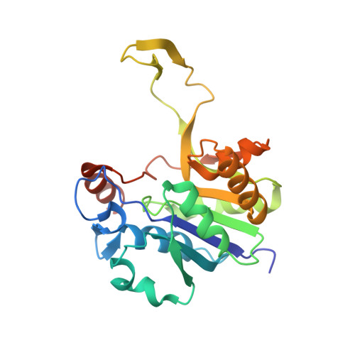
Structure of 4-diphosphocytidyl-2-C- methylerythritol synthetase involved in mevalonate- independent isoprenoid biosynthesis.

Richard, S.B.Bowman, M.E.Kwiatkowski, W.Kang, I.Chow, C.Lillo, A.M.Cane, D.E.Noel, J.P.

(2001) Nat Struct Biol 8: 641-648

  • DOI: https://doi.org/10.1038/89691
  • Primary Citation Related Structures: 
    1I52, 1INI, 1INJ

  • PubMed Abstract: 

    The YgbP protein of Escherichia coli encodes the enzyme 4-diphosphocytidyl-2-C-methylerythritol (CDP-ME) synthetase, a member of the cytidyltransferase family of enzymes. CDP-ME is an intermediate in the mevalonate-independent pathway for isoprenoid biosynthesis in a number of prokaryotic organisms, algae, the plant plastids and the malaria parasite. Because vertebrates synthesize isoprenoid precursors using a mevalonate pathway, CDP-ME synthetase and other enzymes of the mevalonate-independent pathway for isoprenoid production represent attractive targets for the structure-based design of selective antibacterial, herbicidal and antimalarial drugs. The high-resolution structures of E. coli CDP-ME synthetase in the apo form and complexed with both CTP-Mg2+ and CDP-ME-Mg2+ reveal the stereochemical principles underlying both substrate and product recognition as well as catalysis in CDP-ME synthetase. Moreover, these complexes represent the first experimental structures for any cytidyltransferase with both substrates and products bound.


  • Organizational Affiliation
    • Structural Biology Laboratory, The Salk Institute for Biological Studies, 10010 North Torrey Pines Road, La Jolla, California 92037, USA.

Macromolecule Content 

  • Total Structure Weight: 26.32 kDa 
  • Atom Count: 1,853 
  • Modeled Residue Count: 225 
  • Deposited Residue Count: 236 
  • Unique protein chains: 1

Macromolecules

Find similar proteins by:|  3D Structure
Entity ID: 1
MoleculeChains  Sequence LengthOrganismDetailsImage
4-DIPHOSPHOCYTIDYL-2-C-METHYLERYTHRITOL SYNTHETASE236Escherichia coli K-12Mutation(s): 0 
Gene Names: ISPD
EC: 2.7.7 (PDB Primary Data), 2.7.7.60 (UniProt)
UniProt
Find proteins for Q46893 (Escherichia coli (strain K12))
Explore Q46893 
Go to UniProtKB:  Q46893
Entity Groups
Sequence Clusters30% Identity50% Identity70% Identity90% Identity95% Identity100% Identity
UniProt GroupQ46893
Sequence Annotations
Expand
Reference Sequence

Small Molecules

Ligands 2 Unique
IDChains Name / Formula / InChI Key2D Diagram3D Interactions
CDM

Query on CDM


Download:Ideal Coordinates CCD File
C [auth A]4-DIPHOSPHOCYTIDYL-2-C-METHYL-D-ERYTHRITOL
C14 H25 N3 O14 P2
YFAUKWZNPVBCFF-XHIBXCGHSA-N
MG

Query on MG


Download:Ideal Coordinates CCD File
B [auth A]MAGNESIUM ION
Mg
JLVVSXFLKOJNIY-UHFFFAOYSA-N

Experimental Data & Validation

Experimental Data

  • Method: X-RAY DIFFRACTION
  • Resolution: 1.82 Å
  • R-Value Free:  0.273 (Depositor), 0.273 (DCC) 
  • R-Value Work:  0.222 (Depositor), 0.222 (DCC) 
  • R-Value Observed: 0.222 (Depositor) 
Space Group: C 1 2 1
Unit Cell:
Length ( Å )Angle ( ˚ )
a = 129.99α = 90
b = 46.764β = 92.64
c = 38.387γ = 90
Software Package:
Software NamePurpose
DENZOdata reduction
SCALEPACKdata scaling
CNSrefinement
CNSphasing

Structure Validation

View Full Validation Report


Ligand Structure Quality Assessment 


Entry History 

Deposition Data

Revision History  (Full details and data files)

  • Version 1.0: 2001-11-14
    Type: Initial release
  • Version 1.1: 2008-04-27
    Changes: Version format compliance
  • Version 1.2: 2011-07-13
    Changes: Derived calculations, Source and taxonomy, Version format compliance
  • Version 1.3: 2023-08-16
    Changes: Data collection, Database references, Derived calculations, Refinement description