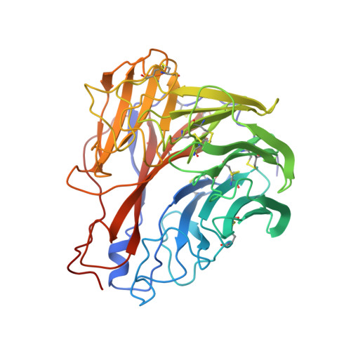
1INH

INFLUENZA A SUBTYPE N2 NEURAMINIDASE COMPLEXED WITH AROMATIC BANA111 INHIBITOR


Experimental Data Snapshot

  • Method: X-RAY DIFFRACTION
  • Resolution: 2.40 Å
  • R-Value Work: 0.184 
  • R-Value Observed: 0.184 

wwPDB Validation   3D Report Full Report


Ligand Structure Quality Assessment 


This is version 2.0 of the entry. See complete history


Literature

Structure-based inhibitors of influenza virus sialidase. A benzoic acid lead with novel interaction.

Singh, S.Jedrzejas, M.J.Air, G.M.Luo, M.Laver, W.G.Brouillette, W.J.

(1995) J Med Chem 38: 3217-3225

  • DOI: https://doi.org/10.1021/jm00017a005
  • Primary Citation of Related Structures:  
    1INF, 1ING, 1INH

  • PubMed Abstract: 

    Influenza virus sialidase is a surface enzyme that is essential for infection of the virus. The catalytic site is highly conserved among all known influenza variants, suggesting that this protein is a suitable target for drug intervention. The most potent known inhibitors are analogs of 2-deoxy-2,3-didehydro-N-acetylneuraminic acid (Neu5Ac2en), particularly the 4-guanidino derivative (4-guanidino-Neu5Ac2en). We utilized the benzene ring of 4-(N-acetylamino)benzoic acids as a cyclic template to substitute for the dihydropyran ring of Neu5Ac2en. In this study several 3-(N-acylamino) derivatives were prepared as potential replacements for the glycerol side chain of Neu5Ac2en, and some were found to interact with the same binding subsite of sialidase. Of greater significance was the observation that the 3-guanidinobenzoic acid derivative (equivalent to the 4-guanidino grouping of 4-guanidino-Neu5Ac2en), the most potent benzoic acid inhibitor of influenza sialidase thus far identified (IC50 = 10 microM), occupied the glycerol-binding subsite on sialidase as opposed to the guanidino-binding subsite. This benzoic acid derivative thus provides a new compound that interacts in a novel manner with the catalytic site of influenza sialidase.


  • Organizational Affiliation

    Department of Chemistry, University of Alabama at Birmingham 35294, USA.


Macromolecules
Find similar proteins by:  (by identity cutoff)  |  3D Structure
Entity ID: 1
MoleculeChains Sequence LengthOrganismDetailsImage
INFLUENZA A SUBTYPE N2 NEURAMINIDASE
A, B
388Influenza A virusMutation(s): 0 
EC: 3.2.1.18
UniProt
Find proteins for P06820 (Influenza A virus (strain A/Tokyo/3/1967 H2N2))
Explore P06820 
Go to UniProtKB:  P06820
Entity Groups  
Sequence Clusters30% Identity50% Identity70% Identity90% Identity95% Identity100% Identity
UniProt GroupP06820
Glycosylation
Glycosylation Sites: 4
Sequence Annotations
Expand
  • Reference Sequence
Oligosaccharides

Help

Entity ID: 2
MoleculeChains Length2D Diagram Glycosylation3D Interactions
2-acetamido-2-deoxy-beta-D-glucopyranose-(1-4)-2-acetamido-2-deoxy-beta-D-glucopyranose
C, F, G, J
2N-Glycosylation
Glycosylation Resources
GlyTouCan:  G42666HT
GlyCosmos:  G42666HT
GlyGen:  G42666HT
Entity ID: 3
MoleculeChains Length2D Diagram Glycosylation3D Interactions
beta-D-mannopyranose-(1-4)-2-acetamido-2-deoxy-beta-D-glucopyranose-(1-4)-[beta-L-fucopyranose-(1-6)]2-acetamido-2-deoxy-beta-D-glucopyranose
D
4N-Glycosylation
Glycosylation Resources
GlyTouCan:  G86566TX
GlyCosmos:  G86566TX
GlyGen:  G86566TX
Entity ID: 4
MoleculeChains Length2D Diagram Glycosylation3D Interactions
beta-D-mannopyranose-(1-2)-alpha-D-mannopyranose-(1-3)-beta-D-mannopyranose-(1-4)-2-acetamido-2-deoxy-beta-D-glucopyranose-(1-4)-[alpha-D-mannopyranose-(1-6)]2-acetamido-2-deoxy-beta-D-glucopyranose
E
6N-Glycosylation
Glycosylation Resources
GlyTouCan:  G64632MT
GlyCosmos:  G64632MT
GlyGen:  G64632MT
Entity ID: 5
MoleculeChains Length2D Diagram Glycosylation3D Interactions
beta-D-mannopyranose-(1-4)-2-acetamido-2-deoxy-beta-D-glucopyranose-(1-4)-[alpha-L-fucopyranose-(1-6)]2-acetamido-2-deoxy-beta-D-glucopyranose
H
4N-Glycosylation
Glycosylation Resources
GlyTouCan:  G32152BH
GlyCosmos:  G32152BH
GlyGen:  G32152BH
Entity ID: 6
MoleculeChains Length2D Diagram Glycosylation3D Interactions
alpha-D-mannopyranose-(1-2)-alpha-D-mannopyranose-(1-3)-beta-D-mannopyranose-(1-4)-2-acetamido-2-deoxy-beta-D-glucopyranose-(1-4)-[alpha-D-mannopyranose-(1-6)]2-acetamido-2-deoxy-beta-D-glucopyranose
I
6N-Glycosylation
Glycosylation Resources
GlyTouCan:  G19158MO
GlyCosmos:  G19158MO
GlyGen:  G19158MO
Small Molecules
Binding Affinity Annotations 
IDSourceBinding Affinity
ST6 PDBBind:  1INH IC50: 5.00e+6 (nM) from 1 assay(s)
Experimental Data & Validation

Experimental Data

  • Method: X-RAY DIFFRACTION
  • Resolution: 2.40 Å
  • R-Value Work: 0.184 
  • R-Value Observed: 0.184 
  • Space Group: C 2 2 21
Unit Cell:
Length ( Å )Angle ( ˚ )
a = 119.74α = 90
b = 141.02β = 90
c = 141.78γ = 90
Software Package:
Software NamePurpose
XENGENdata collection
X-PLORmodel building
X-PLORrefinement
XENGENdata reduction
X-PLORphasing

Structure Validation

View Full Validation Report


Ligand Structure Quality Assessment 


Entry History 

Deposition Data

Revision History  (Full details and data files)

  • Version 1.0: 1996-08-17
    Type: Initial release
  • Version 1.1: 2008-03-24
    Changes: Version format compliance
  • Version 1.2: 2011-07-13
    Changes: Non-polymer description, Version format compliance
  • Version 1.3: 2011-10-26
    Changes: Derived calculations
  • Version 2.0: 2020-07-29
    Type: Remediation
    Reason: Carbohydrate remediation
    Changes: Advisory, Atomic model, Data collection, Database references, Derived calculations, Other, Structure summary