1ILT | pdb_00001ilt

X-RAY STRUCTURE OF INTERLEUKIN-1 RECEPTOR ANTAGONIST AT 2.0 ANGSTROMS RESOLUTION


Experimental Data Snapshot

  • Method: X-RAY DIFFRACTION
  • Resolution: 2.00 Å
  • R-Value Work: 
    0.236 (Depositor) 
  • R-Value Observed: 
    0.236 (Depositor) 

wwPDB Validation 3D Report Full Report

Validation slider image for 1ILT

This is version 1.4 of the entry. See complete history

Literature

X-ray structure of interleukin-1 receptor antagonist at 2.0-A resolution.

Vigers, G.P.Caffes, P.Evans, R.J.Thompson, R.C.Eisenberg, S.P.Brandhuber, B.J.

(1994) J Biological Chem 269: 12874-12879

  • Primary Citation Related Structures: 
    1ILT

  • PubMed Abstract: 

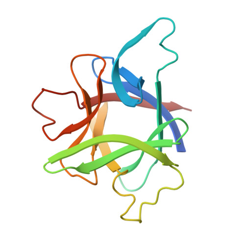
    Interleukin-1 receptor antagonist (IL-1ra) is a natural competitive antagonist of IL-1. In order to further elucidate the mechanism by which IL-1ra binds without activating the IL-1 receptor, we have solved the crystal structure of IL-1ra at 2.0-A resolution. IL-1ra has the same overall beta-trefoil fold as IL-1 alpha and IL-1 beta and has a very similar hydrophobic core. However, there are a number of structural differences between the molecules, including significant differences at the open end of the beta-barrel, which has been identified in IL-1 beta as a receptor binding site.


  • Organizational Affiliation
    • Synergen, Inc., Boulder, Colorado 80301.

Macromolecule Content 

  • Total Structure Weight: 34.29 kDa 
  • Atom Count: 286 
  • Modeled Residue Count: 286 
  • Deposited Residue Count: 304 
  • Unique protein chains: 1

Macromolecules

Find similar proteins by:|  3D Structure
Entity ID: 1
MoleculeChains  Sequence LengthOrganismDetailsImage
INTERLEUKIN-1 RECEPTOR ANTAGONIST
A, B
152Homo sapiensMutation(s): 0 
UniProt & NIH Common Fund Data Resources
Find proteins for P18510 (Homo sapiens)
Explore P18510 
Go to UniProtKB:  P18510
PHAROS:  P18510
GTEx:  ENSG00000136689 
Entity Groups
Sequence Clusters30% Identity50% Identity70% Identity90% Identity95% Identity100% Identity
UniProt GroupP18510
Sequence Annotations
Expand
Reference Sequence

Experimental Data & Validation

Experimental Data

  • Method: X-RAY DIFFRACTION
  • Resolution: 2.00 Å
  • R-Value Work:  0.236 (Depositor) 
  • R-Value Observed: 0.236 (Depositor) 
Space Group: P 43 21 2
Unit Cell:
Length ( Å )Angle ( ˚ )
a = 71.11α = 90
b = 71.11β = 90
c = 112.61γ = 90
Software Package:
Software NamePurpose
X-PLORmodel building
X-PLORrefinement
X-PLORphasing

Structure Validation

View Full Validation Report


Entry History 

Deposition Data

Revision History  (Full details and data files)

  • Version 1.0: 1995-04-01
    Type: Initial release
  • Version 1.1: 2008-03-24
    Changes: Version format compliance
  • Version 1.2: 2011-07-13
    Changes: Version format compliance
  • Version 1.3: 2017-11-29
    Changes: Derived calculations, Other
  • Version 1.4: 2024-02-07
    Changes: Data collection, Database references