1IGW | pdb_00001igw

Crystal Structure of the Isocitrate Lyase from the A219C mutant of Escherichia coli


Experimental Data Snapshot

  • Method: X-RAY DIFFRACTION
  • Resolution: 2.10 Å
  • R-Value Free: 
    0.235 (Depositor) 
  • R-Value Work: 
    0.184 (Depositor), 0.170 (DCC) 
  • R-Value Observed: 
    0.187 (Depositor) 

wwPDB Validation 3D Report Full Report

Validation slider image for 1IGW

This is version 2.0 of the entry. See complete history

Literature

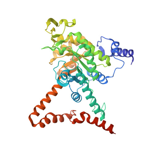
The structure and domain organization of Escherichia coli isocitrate lyase.

Britton, K.L.Abeysinghe, I.S.Baker, P.J.Barynin, V.Diehl, P.Langridge, S.J.McFadden, B.A.Sedelnikova, S.E.Stillman, T.J.Weeradechapon, K.Rice, D.W.

(2001) Acta Crystallogr D Biol Crystallogr 57: 1209-1218

  • DOI: https://doi.org/10.1107/s0907444901008642
  • Primary Citation Related Structures: 
    1IGW

  • PubMed Abstract: 

    Enzymes of the glyoxylate-bypass pathway are potential targets for the control of many human diseases caused by such pathogens as Mycobacteria and Leishmania. Isocitrate lyase catalyses the first committed step in this pathway and the structure of this tetrameric enzyme from Escherichia coli has been determined at 2.1 A resolution. E. coli isocitrate lyase, like the enzyme from other prokaryotes, is located in the cytoplasm, whereas in plants, protozoa, algae and fungi this enzyme is found localized in glyoxysomes. Comparison of the structure of the prokaryotic isocitrate lyase with that from the eukaryote Aspergillus nidulans reveals a different domain structure following the deletion of approximately 100 residues from the larger eukaryotic enzyme. Despite this, the active sites of the prokaryotic and eukaryotic enzymes are very closely related, including the apparent disorder of two equivalent segments of the protein that are known to be involved in a conformational change as part of the enzyme's catalytic cycle.


  • Organizational Affiliation
    • Krebs Institute for Biomolecular Research, Department of Molecular Biology and Biotechnology, The University of Sheffield, Sheffield S10 2TN, England.

Macromolecule Content 

  • Total Structure Weight: 195.06 kDa 
  • Atom Count: 13,435 
  • Modeled Residue Count: 1,628 
  • Deposited Residue Count: 1,736 
  • Unique protein chains: 1

Macromolecules

Find similar proteins by:|  3D Structure
Entity ID: 1
MoleculeChains  Sequence LengthOrganismDetailsImage
Isocitrate lyase
A, B, C, D
434Escherichia coliMutation(s): 1 
Gene Names: aceA
EC: 4.1.3.1
UniProt
Find proteins for P0A9G6 (Escherichia coli (strain K12))
Explore P0A9G6 
Go to UniProtKB:  P0A9G6
Entity Groups
Sequence Clusters30% Identity50% Identity70% Identity90% Identity95% Identity100% Identity
UniProt GroupP0A9G6
Sequence Annotations
Expand
Reference Sequence

Small Molecules

Ligands 3 Unique
IDChains Name / Formula / InChI Key2D Diagram3D Interactions
HG

Query on HG


Download:Ideal Coordinates CCD File
AA [auth D]
BA [auth D]
CA [auth D]
DA [auth D]
E [auth A]
AA [auth D],
BA [auth D],
CA [auth D],
DA [auth D],
E [auth A],
EA [auth D],
F [auth A],
G [auth A],
H [auth A],
I [auth A],
L [auth B],
M [auth B],
N [auth B],
O [auth B],
P [auth B],
S [auth C],
T [auth C],
U [auth C],
V [auth C],
W [auth C],
X [auth C]
MERCURY (II) ION
Hg
BQPIGGFYSBELGY-UHFFFAOYSA-N
PYR

Query on PYR


Download:Ideal Coordinates CCD File
GA [auth D],
K [auth A],
R [auth B],
Z [auth C]
PYRUVIC ACID
C3 H4 O3
LCTONWCANYUPML-UHFFFAOYSA-N
MG

Query on MG


Download:Ideal Coordinates CCD File
FA [auth D],
J [auth A],
Q [auth B],
Y [auth C]
MAGNESIUM ION
Mg
JLVVSXFLKOJNIY-UHFFFAOYSA-N

Experimental Data & Validation

Experimental Data

  • Method: X-RAY DIFFRACTION
  • Resolution: 2.10 Å
  • R-Value Free:  0.235 (Depositor) 
  • R-Value Work:  0.184 (Depositor), 0.170 (DCC) 
  • R-Value Observed: 0.187 (Depositor) 
Space Group: P 32
Unit Cell:
Length ( Å )Angle ( ˚ )
a = 88.65α = 90
b = 88.65β = 90
c = 199.4γ = 120
Software Package:
Software NamePurpose
CCP4model building
TFFCmodel building
DMmodel building
REFMACrefinement
MOSFLMdata reduction
CCP4data scaling
ROTAVATAdata scaling
CCP4phasing
TFFCphasing
DMphasing

Structure Validation

View Full Validation Report


Entry History 

Revision History  (Full details and data files)

  • Version 1.0: 2001-09-05
    Type: Initial release
  • Version 1.1: 2008-04-27
    Changes: Version format compliance
  • Version 1.2: 2011-07-13
    Changes: Version format compliance
  • Version 1.3: 2017-10-04
    Changes: Refinement description
  • Version 1.4: 2021-10-27
    Changes: Database references, Derived calculations
  • Version 2.0: 2023-11-15
    Changes: Atomic model, Data collection, Derived calculations