1IEI | pdb_00001iei

CRYSTAL STRUCTURE OF HUMAN ALDOSE REDUCTASE COMPLEXED WITH THE INHIBITOR ZENARESTAT.


Experimental Data Snapshot

  • Method: X-RAY DIFFRACTION
  • Resolution: 2.50 Å
  • R-Value Free: 
    0.199 (Depositor) 
  • R-Value Work: 
    0.178 (Depositor), 0.167 (DCC) 

wwPDB Validation 3D Report Full Report

Validation slider image for 1IEI

Ligand Structure Quality Assessment 


This is version 1.3 of the entry. See complete history

Literature

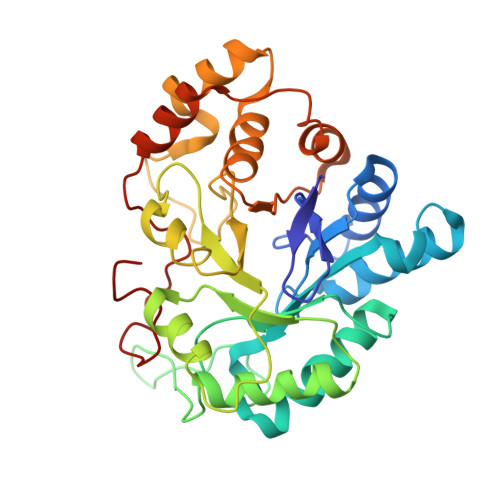
The structure of human recombinant aldose reductase complexed with the potent inhibitor zenarestat.

Kinoshita, T.Miyake, H.Fujii, T.Takakura, S.Goto, T.

(2002) Acta Crystallogr D Biol Crystallogr 58: 622-626

  • DOI: https://doi.org/10.1107/s0907444902002378
  • Primary Citation Related Structures: 
    1IEI

  • PubMed Abstract: 

    The crystal structure of the complex of human recombinant aldose reductase (AR) with zenarestat, one of its potent inhibitors, has been solved at 2.5 A resolution. Zenarestat fits neatly in the hydrophobic active site and induces unique and dramatic conformational changes. For example, the benzene ring of zenarestat occupies a gap in the side chains of Leu300 and Trp111 that interact directly and forms a CH-pi interaction in the native holoenzyme. As a result, the benzene ring of the inhibitor and these side chains form a CH-pi-pi interaction. Such structural information is key to understanding the mode of action of this class of inhibitors and for rational design of better therapeutics.


  • Organizational Affiliation
    • Exploratory Research Laboratories, Fujisawa Pharmaceutical Co. Ltd, 5-2-3, Tokodai, Tsukuba, Ibaraki 300-2698, Japan. takayoshi_kinoshita@po.fujisawa.co.jp

Macromolecule Content 

  • Total Structure Weight: 37.08 kDa 
  • Atom Count: 2,773 
  • Modeled Residue Count: 315 
  • Deposited Residue Count: 316 
  • Unique protein chains: 1

Macromolecules

Find similar proteins by:|  3D Structure
Entity ID: 1
MoleculeChains  Sequence LengthOrganismDetailsImage
ALDOSE REDUCTASE316Homo sapiensMutation(s): 0 
EC: 1.1.1.21 (PDB Primary Data), 1.1.1.372 (UniProt), 1.1.1.300 (UniProt), 1.1.1.54 (UniProt)
UniProt & NIH Common Fund Data Resources
Find proteins for P15121 (Homo sapiens)
Explore P15121 
Go to UniProtKB:  P15121
PHAROS:  P15121
GTEx:  ENSG00000085662 
Entity Groups
Sequence Clusters30% Identity50% Identity70% Identity90% Identity95% Identity100% Identity
UniProt GroupP15121
Sequence Annotations
Expand
Reference Sequence

Small Molecules

Ligands 2 Unique
IDChains Name / Formula / InChI Key2D Diagram3D Interactions
NAP

Query on NAP


Download:Ideal Coordinates CCD File
B [auth A]NADP NICOTINAMIDE-ADENINE-DINUCLEOTIDE PHOSPHATE
C21 H28 N7 O17 P3
XJLXINKUBYWONI-NNYOXOHSSA-N
ZES

Query on ZES


Download:Ideal Coordinates CCD File
C [auth A][3-(4-BROMO-2-FLUORO-BENZYL)-7-CHLORO-2,4-DIOXO-3,4-DIHYDRO-2H-QUINAZOLIN-1-YL]-ACETIC ACID
C17 H11 Br Cl F N2 O4
SXONDGSPUVNZLO-UHFFFAOYSA-N
Binding Affinity Annotations 
IDSourceBinding Affinity
ZES BindingDB:  1IEI IC50: min: 4.4, max: 44 (nM) from 5 assay(s)

Experimental Data & Validation

Experimental Data

  • Method: X-RAY DIFFRACTION
  • Resolution: 2.50 Å
  • R-Value Free:  0.199 (Depositor) 
  • R-Value Work:  0.178 (Depositor), 0.167 (DCC) 
Space Group: P 1
Unit Cell:
Length ( Å )Angle ( ˚ )
a = 40.4α = 76.2
b = 47.97β = 76.7
c = 47.66γ = 67.5
Software Package:
Software NamePurpose
AMoREphasing
X-PLORrefinement
DENZOdata reduction
SCALEPACKdata scaling

Structure Validation

View Full Validation Report


Ligand Structure Quality Assessment 


Entry History 

Deposition Data

Revision History  (Full details and data files)

  • Version 1.0: 2002-04-10
    Type: Initial release
  • Version 1.1: 2008-04-27
    Changes: Version format compliance
  • Version 1.2: 2011-07-13
    Changes: Version format compliance
  • Version 1.3: 2024-03-13
    Changes: Data collection, Database references, Derived calculations