Single mutations at the subunit interface modulate copper reactivity in Photobacterium leiognathi Cu,Zn superoxide dismutase.
Stroppolo, M.E., Pesce, A., D'Orazio, M., O'Neill, P., Bordo, D., Rosano, C., Milani, M., Battistoni, A., Bolognesi, M., Desideri, A.(2001) J Mol Biol 308: 555-563
- PubMed: 11327787 Search on PubMed
- DOI: https://doi.org/10.1006/jmbi.2001.4606
- Primary Citation Related Structures:
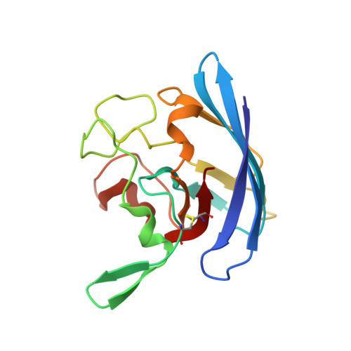
1IB5, 1IBB, 1IBD, 1IBF, 1IBH - PubMed Abstract:
The functional properties and X-ray structures of five mutant forms of Photobacterium leiognathi Cu,Zn superoxide dismutase carrying single mutations at residues located at the dimer association interface have been investigated. When compared to the wild-type enzyme, the three-dimensional structures of the mutants show structural perturbations limited to the proximity of the mutation sites and substantial identity of active site geometry. Nonetheless, the catalytic rates of all mutants, measured at neutral pH and low ionic strength by pulse radiolysis, are higher than that of the wild-type protein. Such enzymatic activity increase is paralleled by enhanced active site accessibility to external chelating agents, which, in the mutated enzyme, remove more readily the active site copper ion. It is concluded that mutations at the prokaryotic Cu,Zn superoxide dismutase subunit interface can transduce dynamical perturbation to the active site region, promoting substrate active site accessibility. Such long-range intramolecular communication effects have not been extensively described before within the Cu,Zn superoxide dismutase homology family.
- Department of Biology and INFM, University of Rome "Tor Vergata", Rome, Via della Ricerca Scientifica, 00133, Italy.
Organizational Affiliation: