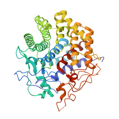
Crystal structure of the cellulase Cel9M enlightens structure/function relationships of the variable catalytic modules in glycoside hydrolases.
Parsiegla, G., Belaich, A., Belaich, J.P., Haser, R.(2002) Biochemistry 41: 11134-11142
- PubMed: 12220178
- DOI: https://doi.org/10.1021/bi025816m
- Primary Citation of Related Structures:
1IA6, 1IA7 - PubMed Abstract:
Cellulases cleave the beta-1.4 glycosidic bond of cellulose. They have been characterized as endo or exo and processive or nonprocessive cellulases according to their action mode on the substrate. Different types of these cellulases may coexist in the same glycoside hydrolase family, which have been classified according to their sequence homology and catalytic mechanism. The bacterium C. celluloyticum produces a set of different cellulases who belong mostly to glycoside hydrolase families 5 and 9. As an adaptation of the organism to different macroscopic substrates organizations and to maximize its cooperative digestion, it is expected that cellulases of these families are active on the various macroscopic organizations of cellulose chains. The nonprocessive cellulase Cel9M is the shortest variant of family 9 cellulases (subgroup 9(C)) which contains only the catalytic module to interact with the substrate. The crystal structures of free native Cel9M and its complex with cellobiose have been solved to 1.8 and 2.0 A resolution, respectively. Other structurally known family 9 cellulases are the nonprocessive endo-cellulase Cel9D from C. thermocellum and the processive endo-cellulase Cel9A from T. fusca, from subgroups 9(B1) and 9(A), respectively, whose catalytic modules are fused to a second domain. These enzymes differ in their activity on substrates with specific macroscopic appearances. The comparison of the catalytic module of Cel9M with the two other known GH family 9 structures may give clues to explain its substrate profile and action mode.
- Institut de Biologie Structurale et Microbiologie, Laboratoire d'Architecture et Fonction des Macromolécules Biologiques, Centre National de la Recherche Scientifique, 31 Chemin Joseph-Aiguier, 13402 Marseille Cedex 20, France.
Organizational Affiliation:


