


The crystal structure of HpcE, a multi-functional enzyme fold
Tame, J.R.H., Namba, K., Dodson, E.J., Roper, D.I.To be published.
Experimental Data Snapshot
wwPDB Validation 3D Report Full Report

Entity ID: 1 | |||||
|---|---|---|---|---|---|
| Molecule | Chains | Sequence Length | Organism | Details | Image |
| 4-HYDROXYPHENYLACETATE DEGRADATION BIFUNCTIONAL ISOMERASE/DECARBOXYLASE | 429 | Escherichia coli | Mutation(s): 0 EC: 5.3.3.10 (PDB Primary Data), 4.1.1.68 (PDB Primary Data) | 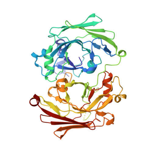 | |
UniProt | |||||
Entity Groups | |||||
| Sequence Clusters | 30% Identity50% Identity70% Identity90% Identity95% Identity100% Identity | ||||
| UniProt Group | P37352 | ||||
Sequence AnnotationsExpand | |||||
Reference Sequence | |||||
| Ligands 1 Unique | |||||
|---|---|---|---|---|---|
| ID | Chains | Name / Formula / InChI Key | 2D Diagram | 3D Interactions | |
| CA Download:Ideal Coordinates CCD File | E [auth A], F [auth B], G [auth C], H [auth D] | CALCIUM ION Ca BHPQYMZQTOCNFJ-UHFFFAOYSA-N |  | ||
| Length ( Å ) | Angle ( ˚ ) |
|---|---|
| a = 126.084 | α = 90 |
| b = 138.201 | β = 90 |
| c = 103.973 | γ = 90 |
| Software Name | Purpose |
|---|---|
| MAR345 | data collection |
| DENZO | data reduction |
| MLPHARE | phasing |
| REFMAC | refinement |
| SCALEPACK | data scaling |


