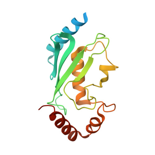
1I7K

CRYSTAL STRUCTURE OF HUMAN MITOTIC-SPECIFIC UBIQUITIN-CONJUGATING ENZYME, UBCH10


Experimental Data Snapshot

  • Method: X-RAY DIFFRACTION
  • Resolution: 1.95 Å
  • R-Value Free: 0.226 
  • R-Value Work: 0.176 

Starting Model: experimental
View more details

wwPDB Validation   3D Report Full Report


This is version 1.4 of the entry. See complete history


Literature

Structural and functional analysis of the human mitotic-specific ubiquitin-conjugating enzyme, UbcH10.

Lin, Y.Hwang, W.C.Basavappa, R.

(2002) J Biol Chem 277: 21913-21921

  • DOI: https://doi.org/10.1074/jbc.M109398200
  • Primary Citation of Related Structures:  
    1I7K

  • PubMed Abstract: 

    Cell cycle progression is controlled at several different junctures by the targeted destruction of cell cycle regulatory proteins. These carefully orchestrated events include the destruction of the securin protein to permit entry into anaphase, and the destruction of cyclin B to permit exit from mitosis. These destruction events are mediated by the ubiquitin/proteasome system. The human ubiquitin-conjugating enzyme, UbcH10, is an essential mediator of the mitotic destruction events. We report here the 1.95-A crystal structure of a mutant UbcH10, in which the active site cysteine has been replaced with a serine. Functional analysis indicates that the mutant is active in accepting ubiquitin, although not as efficiently as wild-type. Examination of the crystal structure reveals that the NH2-terminal extension in UbcH10 is disordered and that a conserved 3(10)-helix places a lysine residue near the active site. Analysis of relevant mutants demonstrates that for ubiquitin-adduct formation the presence or absence of the NH2-terminal extension has little effect, whereas the lysine residue near the active site has significant effect. The structure provides additional insight into UbcH10 function including possible sites of interaction with the anaphase promoting complex/cyclosome and the disposition of a putative destruction box motif in the structure.


  • Organizational Affiliation

    Department of Biochemistry and Biophysics, University of Rochester Medical Center, Rochester, New York 14642, USA.


Macromolecules
Find similar proteins by:  (by identity cutoff)  |  3D Structure
Entity ID: 1
MoleculeChains Sequence LengthOrganismDetailsImage
UBIQUITIN-CONJUGATING ENZYME E2 H10
A, B
179Homo sapiensMutation(s): 1 
Gene Names: UBCH10
EC: 6.3.2.19 (PDB Primary Data), 2.3.2.24 (UniProt), 2.3.2.23 (UniProt)
UniProt & NIH Common Fund Data Resources
Find proteins for O00762 (Homo sapiens)
Explore O00762 
Go to UniProtKB:  O00762
PHAROS:  O00762
GTEx:  ENSG00000175063 
Entity Groups  
Sequence Clusters30% Identity50% Identity70% Identity90% Identity95% Identity100% Identity
UniProt GroupO00762
Sequence Annotations
Expand
  • Reference Sequence
Experimental Data & Validation

Experimental Data

  • Method: X-RAY DIFFRACTION
  • Resolution: 1.95 Å
  • R-Value Free: 0.226 
  • R-Value Work: 0.176 
  • Space Group: P 1
Unit Cell:
Length ( Å )Angle ( ˚ )
a = 40.965α = 62.66
b = 48.371β = 75.26
c = 51.763γ = 81.99
Software Package:
Software NamePurpose
DENZOdata reduction
SCALEPACKdata scaling
AMoREphasing
CNSrefinement
CNSphasing

Structure Validation

View Full Validation Report


Entry History 

Deposition Data

Revision History  (Full details and data files)

  • Version 1.0: 2001-04-04
    Type: Initial release
  • Version 1.1: 2008-04-27
    Changes: Version format compliance
  • Version 1.2: 2011-07-13
    Changes: Version format compliance
  • Version 1.3: 2021-10-27
    Changes: Database references
  • Version 1.4: 2023-08-09
    Changes: Data collection, Refinement description