1I5T | pdb_00001i5t

SOLUTION STRUCTURE OF CYANOFERRICYTOCHROME C


Experimental Data Snapshot

  • Method: SOLUTION NMR
  • Conformers Calculated: 35 
  • Conformers Submitted: 
  • Selection Criteria: all calculated structures submitted 

wwPDB Validation 3D Report Full Report

Validation slider image for 1I5T

This is version 1.4 of the entry. See complete history

Literature

Solution structure of cyanoferricytochrome c: ligand-controlled conformational flexibility and electronic structure of the heme moiety.

Yao, Y.Qian, C.Ye, K.Wang, J.Bai, Z.Tang, W.

(2002) J Biol Inorg Chem 7: 539-547

  • DOI: https://doi.org/10.1007/s00775-001-0334-y
  • Primary Citation Related Structures: 
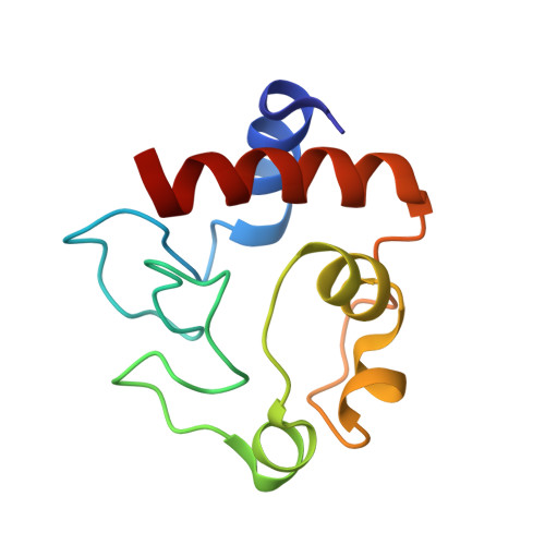
    1I5T

  • PubMed Abstract: 

    The solution structure of cyanoferricytochrome c has been determined using NMR spectroscopy. As a result of including additional constraints derived from pseudocontact shifts, a high-resolution NMR structure was obtained with high accuracy. In order to study the conformational transition between the native protein and its ligand adducts, the present structure was compared with the solution structures of the wild-type cytochrome c and the imidazole-cytochrome c complex. Like the solution structure of imidazole-cytochrome c, the heme crevice is widened by the swinging out of residues 77-85 and a noticeable shift of the 50s helix. However, unlike imidazole, cyanide exerts less significant perturbation on the conformation of the heme cavity, which is revealed by a more compact residue package in the distal pocket. Furthermore, comparison of the solution structure of CN-iso-1Met80Ala cytochrome c with the structure of cyanoferricytochrome c indicated that the binding of cyanide has a different impact on the distal cavity conformation in the two proteins. In addition, the magnetic properties of the present system are discussed and a comprehensive study of the electronic structure of ligand-cytochrome c complexes and the native protein is also described. Electronic supplementary material to this paper can be obtained by using the Springer Link server located at http://dx.doi.org/10.1007/s00775-001-0334-y.


  • Organizational Affiliation
    • State Key Laboratory of Coordination Chemistry, Nanjing University, Nanjing 210093, P.R. China.

Macromolecule Content 

  • Total Structure Weight: 12.37 kDa 
  • Atom Count: 868 
  • Modeled Residue Count: 104 
  • Deposited Residue Count: 104 
  • Unique protein chains: 1

Macromolecules

Find similar proteins by:|  3D Structure
Entity ID: 1
MoleculeChains  Sequence LengthOrganismDetailsImage
CYTOCHROME C104Equus caballusMutation(s): 0 
UniProt
Find proteins for P00004 (Equus caballus)
Explore P00004 
Go to UniProtKB:  P00004
Entity Groups
Sequence Clusters30% Identity50% Identity70% Identity90% Identity95% Identity100% Identity
UniProt GroupP00004
Sequence Annotations
Expand
Reference Sequence

Small Molecules

Ligands 2 Unique
IDChains Name / Formula / InChI Key2D Diagram3D Interactions
HEC

Query on HEC


Download:Ideal Coordinates CCD File
C [auth A]HEME C
C34 H34 Fe N4 O4
HXQIYSLZKNYNMH-LJNAALQVSA-N
CYN

Query on CYN


Download:Ideal Coordinates CCD File
B [auth A]CYANIDE ION
C N
XFXPMWWXUTWYJX-UHFFFAOYSA-N

Experimental Data & Validation

Experimental Data

  • Method: SOLUTION NMR
  • Conformers Calculated: 35 
  • Conformers Submitted: 
  • Selection Criteria: all calculated structures submitted 

Structure Validation

View Full Validation Report


Entry History 

Deposition Data

Revision History  (Full details and data files)

  • Version 1.0: 2001-03-21
    Type: Initial release
  • Version 1.1: 2008-04-27
    Changes: Version format compliance
  • Version 1.2: 2011-07-13
    Changes: Version format compliance
  • Version 1.3: 2022-02-23
    Changes: Data collection, Database references, Derived calculations
  • Version 1.4: 2024-11-20
    Changes: Data collection, Structure summary