1I5J | pdb_00001i5j

NMR STRUCTURE OF HUMAN APOLIPOPROTEIN C-II IN THE PRESENCE OF SDS


Experimental Data Snapshot

  • Method: SOLUTION NMR
  • Conformers Calculated: 100 
  • Conformers Submitted: 25 
  • Selection Criteria: Structures with favourable non-bond energy and minimal constraint violations 

wwPDB Validation 3D Report Full Report

Validation slider image for 1I5J

This is version 1.4 of the entry. See complete history

Literature

NMR structure of human apolipoprotein C-II in the presence of sodium dodecyl sulfate.

MacRaild, C.A.Hatters, D.M.Howlett, G.J.Gooley, P.R.

(2001) Biochemistry 40: 5414-5421

  • DOI: https://doi.org/10.1021/bi002821m
  • Primary Citation Related Structures: 
    1I5J

  • PubMed Abstract: 

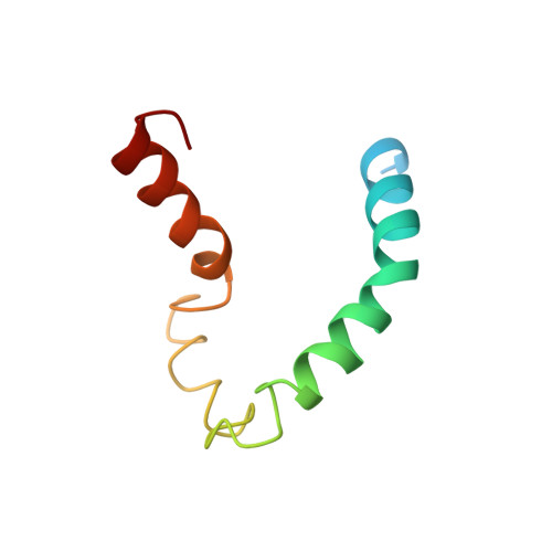
    The structure and protein-detergent interactions of apolipoprotein C-II (apoC-II) in the presence of SDS micelles have been investigated using circular dichroism and heteronuclear NMR techniques applied to (15)N-labeled protein. Micellar SDS, a commonly used mimetic of the lipoprotein surface, inhibits the aggregation of apoC-II and induces a stable structure containing approximately 60% alpha-helix as determined by circular dichroism. NMR reveals the first 12 residues of apoC-II to be structurally heterogeneous and largely disordered, with the rest of the protein forming a predominantly helical structure. Three regions of helical conformation, residues 16-36, 50-56, and 63-77, are well-defined by NMR-derived constraints, with the intervening regions showing more loosely defined helical conformation. The structure of apoC-II is compared to that determined for other apolipoproteins in a similar environment. Our results shed light on the lipid interactions of apoC-II and its mechanism of lipoprotein lipase activation.


  • Organizational Affiliation
    • Russell Grimwade School of Biochemistry and Molecular Biology, University of Melbourne, Gate 12, Royal Parade, Parkville, Victoria 3052, Australia.

Macromolecule Content 

  • Total Structure Weight: 8.92 kDa 
  • Atom Count: 531 
  • Modeled Residue Count: 67 
  • Deposited Residue Count: 79 
  • Unique protein chains: 1

Macromolecules

Find similar proteins by:|  3D Structure
Entity ID: 1
MoleculeChains  Sequence LengthOrganismDetailsImage
APOLIPOPROTEIN CII79Homo sapiensMutation(s): 0 
Membrane Entity: Yes 
UniProt & NIH Common Fund Data Resources
Find proteins for P02655 (Homo sapiens)
Explore P02655 
Go to UniProtKB:  P02655
PHAROS:  P02655
GTEx:  ENSG00000234906 
Entity Groups
Sequence Clusters30% Identity50% Identity70% Identity90% Identity95% Identity100% Identity
UniProt GroupP02655
Sequence Annotations
Expand
Reference Sequence

Experimental Data & Validation

Experimental Data

  • Method: SOLUTION NMR
  • Conformers Calculated: 100 
  • Conformers Submitted: 25 
  • Selection Criteria: Structures with favourable non-bond energy and minimal constraint violations 

Structure Validation

View Full Validation Report


Entry History 

Deposition Data

Revision History  (Full details and data files)

  • Version 1.0: 2001-05-16
    Type: Initial release
  • Version 1.1: 2008-04-27
    Changes: Version format compliance
  • Version 1.2: 2011-07-13
    Changes: Version format compliance
  • Version 1.3: 2022-02-23
    Changes: Database references, Derived calculations
  • Version 1.4: 2024-05-22
    Changes: Data collection