1I1F

Crystal structure of human class i mhc (hla-a2.1) complexed with beta 2-microglobulin and hiv-rt variant peptide i1y


Experimental Data Snapshot

  • Method: X-RAY DIFFRACTION
  • Resolution: 2.80 Å
  • R-Value Free: 0.315 
  • R-Value Work: 0.260 
  • R-Value Observed: 0.274 

wwPDB Validation   3D Report Full Report


This is version 1.3 of the entry. See complete history


Literature

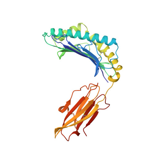
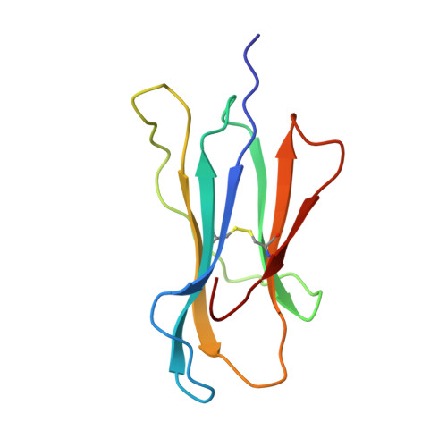
The structural basis for the increased immunogenicity of two HIV-reverse transcriptase peptide variant/class I major histocompatibility complexes.

Kirksey, T.J.Pogue-Caley, R.R.Frelinger, J.A.Collins, E.J.

(1999) J Biol Chem 274: 37259-37264

  • DOI: https://doi.org/10.1074/jbc.274.52.37259
  • Primary Citation of Related Structures:  
    1I1F, 1I1Y

  • PubMed Abstract: 

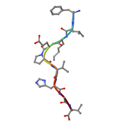
    Designing altered peptide ligands to generate specific immunological reactivity when bound to class I major histocompatibility complexes is important for both therapeutic and prophylactic reasons. We have previously shown that two altered peptides, derived from human immunodeficiency virus (HIV)-reverse transcriptase (RT) residues 309-317, are more immunogenic in vitro than the wild-type peptide. One peptide variant, I1Y, was able to stimulate RT-specific cytotoxic T cells from the blood of three HIV-infected individuals better than the wild-type RT peptide. Both I1Y and I1F peptide variants increase the cell surface half-life of the peptide-class I complex approximately 3-fold over that of the RT peptide but have different immunological activities. These peptides are candidates for the design of vaccines for HIV due to their increased immunogenicity. To understand the basis for the increased cell surface stability compared with wild-type peptide and to understand the differences in T cell recognition between I1Y and I1F, we determined the x-ray crystal structures of the two class I MHC-peptide complexes. These structures indicate that the increased cell surface half-life is due to pi-pi stacking interactions between Trp-167 of HLA-A2.1 and the aromatic P1 residues of I1F and I1Y. Comparison of the structures and modeling potential T cell receptor (TCR) interactions suggests that T cell interactions and immunogenicity are different between I1Y and I1F for two reasons. First, subtle changes in the steric and polar properties of the I1Y peptide affect TCR engagement. Second, water-mediated hydrogen bond interactions between the P1-Tyr and the P4-Glu peptide residues increase peptide side chain rigidity of residues critical for TCR engagement.


  • Organizational Affiliation

    Department of Microbiology, the University of North Carolina, Chapel Hill, North Carolina 27599, USA.


Macromolecules
Find similar proteins by:  (by identity cutoff)  |  3D Structure
Entity ID: 1
MoleculeChains Sequence LengthOrganismDetailsImage
PROTEIN (CLASS I HISTOCOMPATIBILITY ANTIGEN, GOGO-A0201 ALPHA CHAIN)
A, D
275Homo sapiensMutation(s): 0 
UniProt & NIH Common Fund Data Resources
Find proteins for P04439 (Homo sapiens)
Explore P04439 
Go to UniProtKB:  P04439
PHAROS:  P04439
GTEx:  ENSG00000206503 
Entity Groups  
Sequence Clusters30% Identity50% Identity70% Identity90% Identity95% Identity100% Identity
UniProt GroupP04439
Sequence Annotations
Expand
  • Reference Sequence
Find similar proteins by:  (by identity cutoff)  |  3D Structure
Entity ID: 2
MoleculeChains Sequence LengthOrganismDetailsImage
PROTEIN (BETA 2-MICROGLOBULIN)
B, E
100Homo sapiensMutation(s): 0 
UniProt & NIH Common Fund Data Resources
Find proteins for P61769 (Homo sapiens)
Explore P61769 
Go to UniProtKB:  P61769
PHAROS:  P61769
GTEx:  ENSG00000166710 
Entity Groups  
Sequence Clusters30% Identity50% Identity70% Identity90% Identity95% Identity100% Identity
UniProt GroupP61769
Sequence Annotations
Expand
  • Reference Sequence

Find similar proteins by:  Sequence   |   3D Structure  

Entity ID: 3
MoleculeChains Sequence LengthOrganismDetailsImage
PROTEIN (HIV-RT VARIANT PEPTIDE I1F (FLKEPVHGV))
C, F
9N/AMutation(s): 0 
Sequence Annotations
Expand
  • Reference Sequence
Experimental Data & Validation

Experimental Data

  • Method: X-RAY DIFFRACTION
  • Resolution: 2.80 Å
  • R-Value Free: 0.315 
  • R-Value Work: 0.260 
  • R-Value Observed: 0.274 
  • Space Group: P 1
Unit Cell:
Length ( Å )Angle ( ˚ )
a = 49.574α = 82.09
b = 62.97β = 76.47
c = 74.559γ = 77.78
Software Package:
Software NamePurpose
DENZOdata reduction
SCALEPACKdata scaling
AMoREphasing
REFMACrefinement
CCP4phasing

Structure Validation

View Full Validation Report


Entry History 

Deposition Data

Revision History  (Full details and data files)

  • Version 1.0: 2000-01-12
    Type: Initial release
  • Version 1.1: 2008-04-26
    Changes: Version format compliance
  • Version 1.2: 2011-07-13
    Changes: Version format compliance
  • Version 1.3: 2023-08-09
    Changes: Data collection, Database references, Refinement description