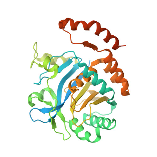
Crystallographic structure and functional interpretation of the cytoplasmic domain of erythrocyte membrane band 3.
Zhang, D., Kiyatkin, A., Bolin, J.T., Low, P.S.(2000) Blood 96: 2925-2933
- PubMed: 11049968 Search on PubMed
- Primary Citation Related Structures:
1HYN - PubMed Abstract:
The red blood cell membrane (RBCM) is a primary model for animal cell plasma membranes. One of its major organizing centers is the cytoplasmic domain of band 3 (cdb3), which links multiple proteins to the membrane. Included among its peripheral protein ligands are ankyrin (the major bridge to the spectrin-actin skeleton), protein 4. 1, protein 4.2, aldolase, glyceraldehyde-3-phosphate dehydrogenase, phosphofructokinase, deoxyhemoglobin, p72syk protein tyrosine kinase, and hemichromes. The crystal structure of cdb3 is reported at 0.26 nm (2.6 A) resolution. A tight symmetric dimer is formed by cdb3; it is stabilized by interlocked dimerization arms contributed by both monomers. Each subunit also includes a larger peripheral protein binding domain with an alpha(+) beta-fold. The binding sites of several peripheral proteins are localized in the structure, and the nature of the major conformational change that regulates membrane-skeletal interactions is evaluated. An improved structural definition of the protein network at the inner surface of the RBCM is now possible.
- Departments of Chemistry and Biological Sciences, Purdue University, West Lafayette, IN 47907-1392, USA.
Organizational Affiliation: