1HWW

GOLGI ALPHA-MANNOSIDASE II IN COMPLEX WITH SWAINSONINE


Experimental Data Snapshot

  • Method: X-RAY DIFFRACTION
  • Resolution: 1.87 Å
  • R-Value Free: 0.209 
  • R-Value Work: 0.181 

wwPDB Validation   3D Report Full Report


Ligand Structure Quality Assessment 


This is version 1.4 of the entry. See complete history


Literature

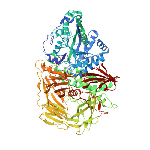
Structure of Golgi alpha-mannosidase II: a target for inhibition of growth and metastasis of cancer cells.

van den Elsen, J.M.Kuntz, D.A.Rose, D.R.

(2001) EMBO J 20: 3008-3017

  • DOI: https://doi.org/10.1093/emboj/20.12.3008
  • Primary Citation of Related Structures:  
    1HTY, 1HWW, 1HXK

  • PubMed Abstract: 
  • Golgi alpha-mannosidase II, a key enzyme in N-glycan processing, is a target in the development of anti- cancer therapies. The crystal structure of Drosophila Golgi alpha-mannosidase II in the absence and presence of the anti-cancer agent swainsonine and the inhibitor deoxymannojirimycin reveals a novel protein fold with an active site zinc intricately involved both in the substrate specificity of the enzyme and directly in the catalytic mechanism ...

    Golgi alpha-mannosidase II, a key enzyme in N-glycan processing, is a target in the development of anti- cancer therapies. The crystal structure of Drosophila Golgi alpha-mannosidase II in the absence and presence of the anti-cancer agent swainsonine and the inhibitor deoxymannojirimycin reveals a novel protein fold with an active site zinc intricately involved both in the substrate specificity of the enzyme and directly in the catalytic mechanism. Identification of a putative GlcNAc binding pocket in the vicinity of the active site cavity provides a model for the binding of the GlcNAcMan(5)GlcNAc(2) substrate and the consecutive hydrolysis of the alpha1,6- and alpha1,3-linked mannose residues. The enzyme-inhibitor interactions observed provide insight into the catalytic mechanism, opening the door to the design of novel inhibitors of alpha-mannosidase II.


    Organizational Affiliation

    Ontario Cancer Institute and Department of Medical Biophysics, University of Toronto, 610 University Avenue, Toronto, Ontario, Canada.


Macromolecules
Find similar proteins by:  (by identity cutoff)  |  3D Structure
Entity ID: 1
MoleculeChainsSequence LengthOrganismDetailsImage
ALPHA-MANNOSIDASE IIA1,015Drosophila melanogasterMutation(s): 0 
Gene Names: alpha-Man-IIaGmIICG18802
EC: 3.2.1.114
UniProt
Find proteins for Q24451 (Drosophila melanogaster)
Explore Q24451 
Go to UniProtKB:  Q24451
Entity Groups  
Sequence Clusters30% Identity50% Identity70% Identity90% Identity95% Identity100% Identity
UniProt GroupQ24451
Protein Feature View
Expand
  • Reference Sequence
Small Molecules
Ligands 4 Unique
IDChainsName / Formula / InChI Key2D Diagram3D Interactions
NAG
Query on NAG

Download Ideal Coordinates CCD File 
B [auth A]2-acetamido-2-deoxy-beta-D-glucopyranose
C8 H15 N O6
OVRNDRQMDRJTHS-FMDGEEDCSA-N
 Ligand Interaction
SWA
Query on SWA

Download Ideal Coordinates CCD File 
D [auth A]1S-8AB-OCTAHYDRO-INDOLIZIDINE-1A,2A,8B-TRIOL
C8 H15 N O3
FXUAIOOAOAVCGD-WCTZXXKLSA-N
 Ligand Interaction
MRD
Query on MRD

Download Ideal Coordinates CCD File 
E [auth A](4R)-2-METHYLPENTANE-2,4-DIOL
C6 H14 O2
SVTBMSDMJJWYQN-RXMQYKEDSA-N
 Ligand Interaction
ZN
Query on ZN

Download Ideal Coordinates CCD File 
C [auth A]ZINC ION
Zn
PTFCDOFLOPIGGS-UHFFFAOYSA-N
 Ligand Interaction
Binding Affinity Annotations 
IDSourceBinding Affinity
SWA BindingDB:  1HWW Ki: 3 (nM) from 1 assay(s)
IC50: 37 (nM) from 1 assay(s)
Binding MOAD:  1HWW IC50: 20 (nM) from 1 assay(s)
PDBBind:  1HWW IC50: 20 (nM) from 1 assay(s)
Experimental Data & Validation

Experimental Data

  • Method: X-RAY DIFFRACTION
  • Resolution: 1.87 Å
  • R-Value Free: 0.209 
  • R-Value Work: 0.181 
  • Space Group: P 21 21 21
Unit Cell:
Length ( Å )Angle ( ˚ )
a = 68.902α = 90
b = 110.015β = 90
c = 138.472γ = 90
Software Package:
Software NamePurpose
CNSrefinement
DENZOdata reduction
SCALEPACKdata scaling
CNSphasing

Structure Validation

View Full Validation Report


Ligand Structure Quality Assessment 


Entry History 

Deposition Data

Revision History  (Full details and data files)

  • Version 1.0: 2002-01-10
    Type: Initial release
  • Version 1.1: 2008-04-27
    Changes: Version format compliance
  • Version 1.2: 2011-07-13
    Changes: Non-polymer description, Version format compliance
  • Version 1.3: 2018-04-04
    Changes: Data collection
  • Version 1.4: 2020-07-29
    Type: Remediation
    Reason: Carbohydrate remediation
    Changes: Data collection, Derived calculations, Structure summary