1HUG | pdb_00001hug

Differences in anionic inhibition of Human Carbonic Anhydrase I revealed from the structures of iodide and gold cyanide inhibitor complexes


Experimental Data Snapshot

  • Method: X-RAY DIFFRACTION
  • Resolution: 2.00 Å
  • R-Value Work: 
    0.166 (DCC) 
  • R-Value Observed: 
    0.171 (Depositor) 

wwPDB Validation 3D Report Full Report

Validation slider image for 1HUG

This is version 1.4 of the entry. See complete history

Literature

Differences in anionic inhibition of human carbonic anhydrase I revealed from the structures of iodide and gold cyanide inhibitor complexes.

Kumar, V.Kannan, K.K.Sathyamurthi, P.

(1994) Acta Crystallogr D Biol Crystallogr 50: 731-738

  • DOI: https://doi.org/10.1107/S0907444994001873
  • Primary Citation Related Structures: 
    1HUG, 1HUH

  • PubMed Abstract: 

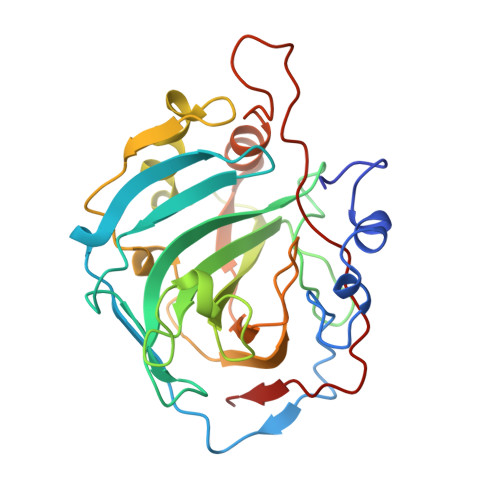
    The crystal structures of two anionic inhibitor complexes of human carbonic anhydrase I (HCAI), namely, HCAI-iodide and HCAI-Au(CN)(2)(-), have been refined by the restrained least-squares method at 2.2 and 2 A nominal resolution, respectively, with good stereochemistry for the final models. The R values have improved from 30.3 to 16.6% for HCAI-iodide and from 28.8 to 17.1% for HCAI-Au(CN)(2)(-). The sites of inhibitor binding as elucidated are totally different in the two structures. The iodide anion replaces the zinc-bound H(2)O/OH(-) ligand and renders the enzyme inactive. This result confirms that the zinc-bound H(2)O/OH(-) is the activity-linked group in carbonic anhydrase enzymes. Au(CN)(2)(-) binds at a different and new site near the zinc ion, without liganding to the metal. The N atom of Au(CN)(2)(-) is within hydrogen-bonding distance of the zinc-bound H(2)O/OH(-) group which shifts by about 0.4 A away from the zinc ion in relation to its position in the native HCAI. It is proposed that the presence of the inhibitor Au(CN)(2)(-) results in a conformational reorientation of the activity-linked group, due to hydrogen-bond formation with the inhibitor, which in turn sterically hinders the binding of the substrate CO(2) molecule in the active site, leading to the inhibition of HCAI enzyme.


  • Organizational Affiliation
    • Solid State Physics Division, Bhabha Atomic Research Centre, Bombay, India.

Macromolecule Content 

  • Total Structure Weight: 29.84 kDa 
  • Atom Count: 2,250 
  • Modeled Residue Count: 256 
  • Deposited Residue Count: 260 
  • Unique protein chains: 1

Macromolecules

Find similar proteins by:|  3D Structure
Entity ID: 1
MoleculeChains  Sequence LengthOrganismDetailsImage
CARBONIC ANHYDRASE I260Homo sapiensMutation(s): 0 
EC: 4.2.1.1 (PDB Primary Data), 4.2.1.69 (UniProt)
UniProt & NIH Common Fund Data Resources
Find proteins for P00915 (Homo sapiens)
Explore P00915 
Go to UniProtKB:  P00915
PHAROS:  P00915
GTEx:  ENSG00000133742 
Entity Groups
Sequence Clusters30% Identity50% Identity70% Identity90% Identity95% Identity100% Identity
UniProt GroupP00915
Sequence Annotations
Expand
Reference Sequence

Experimental Data & Validation

Experimental Data

  • Method: X-RAY DIFFRACTION
  • Resolution: 2.00 Å
  • R-Value Work:  0.166 (DCC) 
  • R-Value Observed: 0.171 (Depositor) 
Space Group: P 21 21 21
Unit Cell:
Length ( Å )Angle ( ˚ )
a = 81.8α = 90
b = 75.2β = 90
c = 37.1γ = 90
Software Package:
Software NamePurpose
PROLSQrefinement

Structure Validation

View Full Validation Report


Entry History 

Deposition Data

Revision History  (Full details and data files)

  • Version 1.0: 1994-04-30
    Type: Initial release
  • Version 1.1: 2008-03-03
    Changes: Version format compliance
  • Version 1.2: 2011-07-13
    Changes: Version format compliance
  • Version 1.3: 2017-11-29
    Changes: Derived calculations, Other
  • Version 1.4: 2024-02-07
    Changes: Data collection, Database references, Derived calculations