1HPY | pdb_00001hpy

THE SOLUTION STRUCTURE OF HUMAN PARATHYROID HORMONE FRAGMENT 1-34 IN 20% TRIFLUORETHANOL, NMR, 10 STRUCTURES


Experimental Data Snapshot

  • Method: SOLUTION NMR
  • Conformers Calculated: 30 
  • Conformers Submitted: 10 
  • Selection Criteria: ENERGY, AGREEMENT WITH EXPERIMENTAL DATA 

wwPDB Validation 3D Report Full Report

Validation slider image for 1HPY

This is version 1.4 of the entry. See complete history

Literature

Solution structures of human parathyroid hormone fragments hPTH(1-34) and hPTH(1-39) and bovine parathyroid hormone fragment bPTH(1-37).

Marx, U.C.Adermann, K.Bayer, P.Forssmann, W.G.Rosch, P.

(2000) Biochem Biophys Res Commun 267: 213-220

  • DOI: https://doi.org/10.1006/bbrc.1999.1958
  • Primary Citation Related Structures: 
    1BWX, 1HPY, 1ZWA, 1ZWC

  • PubMed Abstract: 

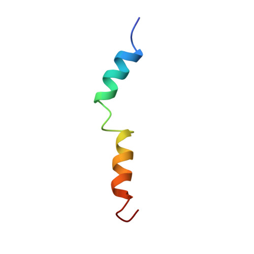
    Parathyroid hormone (PTH) is involved in regulation of the calcium level in blood and has an influence on bone metabolism, thus playing a role in osteoporosis therapy. In this study, the structures of the human PTH fragments (1-34) and (1-39) as well as bovine PTH(1-37) in aqueous buffer solution under near physiological conditions were determined using two-dimensional nuclear magnetic resonance spectroscopy. The overall structure of the first 34 amino acids of these three peptides is virtually identical, exhibiting a short NH(2)-terminal and a longer COOH-terminal helix as well as a defined loop region from His14 to Ser17, stabilized by hydrophobic interactions. bPTH(1-37), which has a higher biological activity, shows a better-defined NH(2)-terminal part. In contrast to NH(2)-terminal truncations, which cause destabilization of helical structure, neither COOH-terminal truncation nor elongation significantly influences the secondary structure. Furthermore, we investigated the structure of hPTH(1-34) in 20% trifluoroethanol solution. In addition to its helix-stabilizing effect, trifluorethanol causes the loss of tertiary hydrophobic interactions.


  • Organizational Affiliation
    • Lehrstuhl für Biopolymere, Universität Bayreuth, Bayreuth, D-95440, Germany. ute.marxf@uni-bayreuth.de

Macromolecule Content 

  • Total Structure Weight: 4.13 kDa 
  • Atom Count: 289 
  • Modeled Residue Count: 34 
  • Deposited Residue Count: 34 
  • Unique protein chains: 1

Macromolecules

Find similar proteins by:|  3D Structure
Entity ID: 1
MoleculeChains  Sequence LengthOrganismDetailsImage
PARATHYROID HORMONE34Homo sapiensMutation(s): 0 
UniProt & NIH Common Fund Data Resources
Find proteins for P01270 (Homo sapiens)
Explore P01270 
Go to UniProtKB:  P01270
PHAROS:  P01270
GTEx:  ENSG00000152266 
Entity Groups
Sequence Clusters30% Identity50% Identity70% Identity90% Identity95% Identity100% Identity
UniProt GroupP01270
Sequence Annotations
Expand
Reference Sequence

Experimental Data & Validation

Experimental Data

  • Method: SOLUTION NMR
  • Conformers Calculated: 30 
  • Conformers Submitted: 10 
  • Selection Criteria: ENERGY, AGREEMENT WITH EXPERIMENTAL DATA 

Structure Validation

View Full Validation Report


Entry History 

Deposition Data

Revision History  (Full details and data files)

  • Version 1.0: 2000-01-14
    Type: Initial release
  • Version 1.1: 2008-03-24
    Changes: Version format compliance
  • Version 1.2: 2011-07-13
    Changes: Version format compliance
  • Version 1.3: 2022-02-23
    Changes: Database references, Derived calculations, Other
  • Version 1.4: 2024-05-22
    Changes: Data collection