1HMC

THREE-DIMENSIONAL STRUCTURE OF DIMERIC HUMAN RECOMBINANT MACROPHAGE COLONY STIMULATING FACTOR


Experimental Data Snapshot

  • Method: X-RAY DIFFRACTION
  • Resolution: 2.50 Å
  • R-Value Work: 0.197 
  • R-Value Observed: 0.197 

wwPDB Validation   3D Report Full Report


This is version 1.3 of the entry. See complete history


Literature

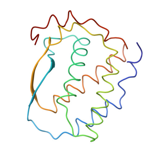
Three-dimensional structure of dimeric human recombinant macrophage colony-stimulating factor.

Pandit, J.Bohm, A.Jancarik, J.Halenbeck, R.Koths, K.Kim, S.H.

(1992) Science 258: 1358-1362

  • DOI: https://doi.org/10.1126/science.1455231
  • Primary Citation of Related Structures:  
    1HMC

  • PubMed Abstract: 

    Macrophage colony-stimulating factor (M-CSF) triggers the development of cells of the monocyte-macrophage lineage and has a variety of stimulatory effects on mature cells of this class. The biologically active form of M-CSF is a disulfide-linked dimer that activates an intrinsic tyrosine kinase activity on the M-CSF receptor by inducing dimerization of the receptor molecules. The structure of a recombinant human M-CSF dimer, determined at 2.5 angstroms by x-ray crystallography, contains two bundles of four alpha helices laid end-to-end, with an interchain disulfide bond. Individual monomers of M-CSF show a close structural similarity to the cytokines granulocyte-macrophage colony-stimulating factor and human growth hormone. Both of these cytokines are monomeric in their active form, and their specific receptors lack intrinsic tyrosine kinase activity. The similarity of these structures suggests that the receptor binding determinants for all three cytokines may be similar.


  • Organizational Affiliation

    Structural Biology Division, Lawrence Berkeley Laboratory, Berkeley, CA 94720.


Macromolecules
Find similar proteins by:  (by identity cutoff)  |  3D Structure
Entity ID: 1
MoleculeChains Sequence LengthOrganismDetailsImage
HUMAN MACROPHAGE COLONY STIMULATING FACTOR
A, B
148Homo sapiensMutation(s): 0 
UniProt & NIH Common Fund Data Resources
Find proteins for P09603 (Homo sapiens)
Explore P09603 
Go to UniProtKB:  P09603
PHAROS:  P09603
GTEx:  ENSG00000184371 
Entity Groups  
Sequence Clusters30% Identity50% Identity70% Identity90% Identity95% Identity100% Identity
UniProt GroupP09603
Sequence Annotations
Expand
  • Reference Sequence
Experimental Data & Validation

Experimental Data

  • Method: X-RAY DIFFRACTION
  • Resolution: 2.50 Å
  • R-Value Work: 0.197 
  • R-Value Observed: 0.197 
  • Space Group: P 21 21 21
Unit Cell:
Length ( Å )Angle ( ˚ )
a = 33.37α = 90
b = 64.75β = 90
c = 157.87γ = 90
Software Package:
Software NamePurpose
X-PLORmodel building
X-PLORrefinement
X-PLORphasing

Structure Validation

View Full Validation Report


Entry History 

Deposition Data

Revision History  (Full details and data files)

  • Version 1.0: 1994-04-30
    Type: Initial release
  • Version 1.1: 2008-03-24
    Changes: Version format compliance
  • Version 1.2: 2011-07-13
    Changes: Version format compliance
  • Version 1.3: 2024-02-07
    Changes: Data collection, Database references, Other