Structure Determination of the Extracellular Xylanase from Geobacillus Stearothermophilus by Selenomethionyl MAD Phasing
Teplitsky, A., Mechaly, A., Stojanoff, V., Sainz, G., Golan, G., Feinberg, H., Gilboa, R., Reiland, V., Zolotnitsky, G., Shallom, D., Thompson, A., Shoham, Y., Shoham, G.(2004) Acta Crystallogr D Biol Crystallogr 60: 836
- PubMed: 15103129 Search on PubMed
- DOI: https://doi.org/10.1107/S0907444904004123
- Primary Citation Related Structures:
1HIZ - PubMed Abstract:
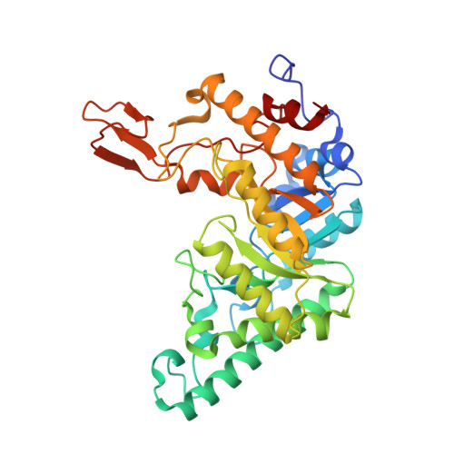
Xylanases are hemicellulases that hydrolyze the internal beta-1,4-glycoside bonds of xylan. The extracellular thermostable endo-1,4-beta-xylanase (EC 3.2.1.8; XT6) produced by the thermophilic bacterium Geobacillus stearothermophilus T-6 was shown to bleach pulp optimally at pH 9 and 338 K and was successfully used in a large-scale biobleaching mill trial. The xylanase gene was cloned and sequenced. The mature enzyme consists of 379 amino acids, with a calculated molecular weight of 43 808 Da and a pI of 9.0. Crystallographic studies of XT6 were performed in order to study the mechanism of catalysis and to provide a structural basis for the rational introduction of enhanced thermostability by site-specific mutagenesis. XT6 was crystallized in the primitive trigonal space group P3(2)21, with unit-cell parameters a = b = 112.9, c = 122.7 A. A full diffraction data set for wild-type XT6 has been measured to 2.4 A resolution on flash-frozen crystals using synchrotron radiation. A fully exchanged selenomethionyl XT6 derivative (containing eight Se atoms per XT6 molecule) was also prepared and crystallized in an isomorphous crystal form, providing full selenium MAD data at three wavelengths and enabling phase solution and structure determination. The structure of wild-type XT6 was refined at 2.4 A resolution to a final R factor of 15.6% and an R(free) of 18.6%. The structure demonstrates that XT6 is made up of an eightfold TIM-barrel containing a deep active-site groove, consistent with its 'endo' mode of action. The two essential catalytic carboxylic residues (Glu159 and Glu265) are located at the active site within 5.5 A of each other, as expected for 'retaining' glycoside hydrolases. A unique subdomain was identified in the carboxy-terminal part of the enzyme and was suggested to have a role in xylan binding. The three-dimensional structure of XT6 is of great interest since it provides a favourable starting point for the rational improvement of its already high thermal and pH stabilities, which are required for a number of biotechnological and industrial applications.
- Department of Inorganic Chemistry, The Hebrew University of Jerusalem, Jerusalem 91904, Israel.
Organizational Affiliation: