1HHJ

THE ANTIGENIC IDENTITY OF PEPTIDE(SLASH)MHC COMPLEXES: A COMPARISON OF THE CONFORMATION OF FIVE PEPTIDES PRESENTED BY HLA-A2


Experimental Data Snapshot

  • Method: X-RAY DIFFRACTION
  • Resolution: 2.50 Å
  • R-Value Work: 0.278 
  • R-Value Observed: 0.278 

wwPDB Validation   3D Report Full Report


This is version 1.3 of the entry. See complete history


Literature

The antigenic identity of peptide-MHC complexes: a comparison of the conformations of five viral peptides presented by HLA-A2.

Madden, D.R.Garboczi, D.N.Wiley, D.C.

(1993) Cell 75: 693-708

  • DOI: https://doi.org/10.1016/0092-8674(93)90490-h
  • Primary Citation of Related Structures:  
    1HHG, 1HHH, 1HHI, 1HHJ, 1HHK

  • PubMed Abstract: 

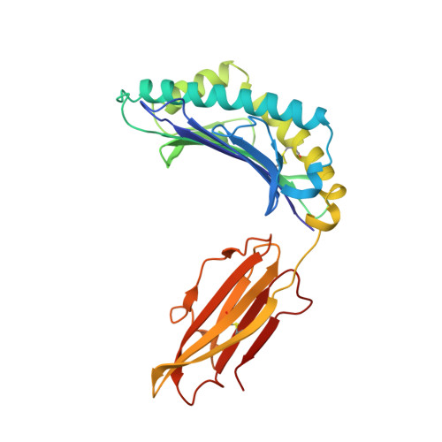
    Complexes of five peptides (from HIV-1, influenza A virus, HTLV-1, and hepatitis B virus proteins) bound to the human class I MHC molecule HLA-A2 have been studied by X-ray crystallography. While the peptide termini and their second and C-terminal anchor side chains are bound similarly in all five cases, the main chain and side chain conformations of each peptide are strikingly different in the center of the binding site, and these differences are accessible to direct TCR recognition. Each of the central peptide residues is seen to point up for some bound peptides, but down or sideways for others. Thus, although fixed at its ends, the structure of an MHC-bound peptide appears to be a highly complex function of its entire sequence, potentially sensitive to even small sequence differences. In contrast, MHC structural variation is relatively limited. These results offer a structural framework for understanding the role of nonanchor peptide side chains in both peptide-MHC binding affinity and TCR recognition.


  • Organizational Affiliation

    Department of Biochemistry and Molecular Biology, Howard Hughes Medical Institute, Harvard University, Cambridge, Massachusetts 02138.


Macromolecules
Find similar proteins by:  (by identity cutoff)  |  3D Structure
Entity ID: 1
MoleculeChains Sequence LengthOrganismDetailsImage
CLASS I HISTOCOMPATIBILITY ANTIGEN (HLA-A*0201) (ALPHA CHAIN)
A, D
275Homo sapiensMutation(s): 0 
Gene Names: BETA-2-MICROGLOBULIN
UniProt & NIH Common Fund Data Resources
Find proteins for P04439 (Homo sapiens)
Go to UniProtKB:  P04439
PHAROS:  P04439
GTEx:  ENSG00000206503 
Sequence Annotations
Expand
  • Reference Sequence
Find similar proteins by:  (by identity cutoff)  |  3D Structure
Entity ID: 2
MoleculeChains Sequence LengthOrganismDetailsImage
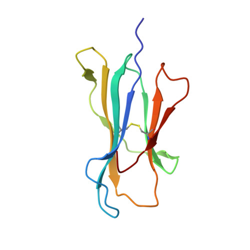
BETA 2-MICROGLOBULIN
B, E
100Homo sapiensMutation(s): 0 
Gene Names: BETA-2-MICROGLOBULIN
UniProt & NIH Common Fund Data Resources
Find proteins for P61769 (Homo sapiens)
Go to UniProtKB:  P61769
PHAROS:  P61769
GTEx:  ENSG00000166710 
Sequence Annotations
Expand
  • Reference Sequence

Find similar proteins by:  Sequence   |   3D Structure  

Entity ID: 3
MoleculeChains Sequence LengthOrganismDetailsImage
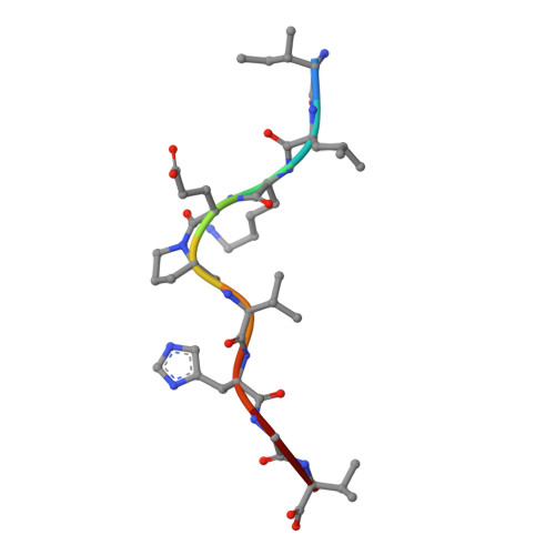
HIV-1 REVERSE TRANSCRIPTASE (RESIDUES 309-317)
C, F
9Human immunodeficiency virus 1Mutation(s): 0 
UniProt
Find proteins for P04588 (Human immunodeficiency virus type 1 group M subtype A (isolate MAL))
Go to UniProtKB:  P04588
Sequence Annotations
Expand
  • Reference Sequence
Experimental Data & Validation

Experimental Data

  • Method: X-RAY DIFFRACTION
  • Resolution: 2.50 Å
  • R-Value Work: 0.278 
  • R-Value Observed: 0.278 
  • Space Group: P 1
Unit Cell:
Length ( Å )Angle ( ˚ )
a = 50.37α = 81.53
b = 63.6β = 75.72
c = 74.75γ = 77.48
Software Package:
Software NamePurpose
X-PLORmodel building
X-PLORrefinement
X-PLORphasing

Structure Validation

View Full Validation Report


Entry History 

Deposition Data

Revision History  (Full details and data files)

  • Version 1.0: 1993-10-31
    Type: Initial release
  • Version 1.1: 2008-03-24
    Changes: Version format compliance
  • Version 1.2: 2011-07-13
    Changes: Version format compliance
  • Version 1.3: 2024-06-05
    Changes: Data collection, Database references, Other