Investigating the structural determinants of the p21-like triphosphate and Mg2+ binding site.
Cronet, P., Bellsolell, L., Sander, C., Coll, M., Serrano, L.(1995) J Mol Biol 249: 654-664
- PubMed: 7783218 Search on PubMed
- DOI: https://doi.org/10.1006/jmbi.1995.0326
- Primary Citation Related Structures:
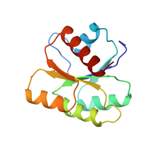
1HEY - PubMed Abstract:
Amongst the superfamily of nucleotide binding proteins, the classical mononucleotide binding fold (CMBF), is the one that has been best characterized structurally. The common denominator of all the members is the triphosphate/Mg2+ binding site, whose signature has been recognized as two structurally conserved stretches of residues: the Kinase 1 and 2 motifs that participate in triphosphate and Mg2+ binding, respectively. The Kinase 1 motif is borne by a loop (the P-loop), whose structure is conserved throughout the whole CMBF family. The low sequence similarity between the different members raises questions about which interactions are responsible for the active structure of the P-loop. What are the minimal requirements for the active structure of the P-loop? Why is the P-loop structure conserved despite the diverse environments in which it is found? To address this question, we have engineered the Kinase 1 and 2 motifs into a protein that has the CMBF and no nucleotide binding activity, the chemotactic protein from Escherichia coli, CheY. The mutant does not exhibit any triphosphate/Mg2+ binding activity. The crystal structure of the mutant reveals that the engineered P-loop is in a different conformation than that found in the CMBF. This demonstrates that the native structure of the P-loop requires external interactions with the rest of the protein. On the basis of an analysis of the conserved tertiary contacts of the P-loop in the mononucleotide binding superfamily, we propose a set of residues that could play an important role in the acquisition of the active structure of the P-loop.
- European Molecular Biology Laboratory, Heidelberg, Germany.
Organizational Affiliation: