1H9R | pdb_00001h9r

Tungstate bound complex Dimop domain of ModE from E.coli


Experimental Data Snapshot

  • Method: X-RAY DIFFRACTION
  • Resolution: 1.90 Å
  • R-Value Free: 
    0.225 (Depositor) 
  • R-Value Work: 
    0.170 (Depositor) 

Starting Model: experimental
View more details

wwPDB Validation 3D Report Full Report

Validation slider image for 1H9R

This is version 1.3 of the entry. See complete history

Literature

Oxyanion Binding Alters Conformational and Quaternary Structure of the C-Terminal Domain of the Transcriptional Regulator Mode; Implications for Molybdate-Dependant Regulation, Signalling, Storage and Transport

Gourley, D.G.Schuttelkopf, A.W.Anderson, L.A.Price, N.C.Boxer, D.H.Hunter, W.N.

(2001) J Biological Chem 276: 20641

  • DOI: https://doi.org/10.1074/jbc.M100919200
  • Primary Citation Related Structures: 
    1H9R, 1H9S

  • PubMed Abstract: 

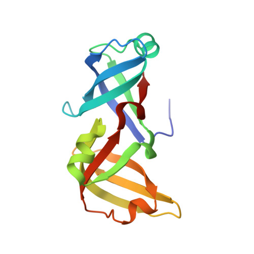
    The molybdate-dependent transcriptional regulator ModE of Escherichia coli functions as a sensor of intracellular molybdate concentration and a regulator for the transcription of several operons that control the uptake and utilization of molybdenum. We present two high-resolution crystal structures of the C-terminal oxyanion-binding domain in complex with molybdate and tungstate. The ligands bind between subunits at the dimerization interface, and analysis reveals that oxyanion selectivity is determined primarily by size. The relevance of the structures is indicated by fluorescence measurements, which show that the oxyanion binding properties of the C-terminal domain of ModE are similar to those of the full-length protein. Comparisons with the apoprotein structure have identified structural rearrangements that occur on binding oxyanion. This molybdate-dependent conformational switch promotes a change in shape and alterations to the surface of the protein and may provide the signal for recruitment of other proteins to construct the machinery for transcription. Sequence and structure-based comparisons lead to a classification of molybdate-binding proteins.


  • Organizational Affiliation
    • Wellcome Trust Biocentre, University of Dundee, Dundee, DD1 5EH, United Kingdom.

Macromolecule Content 

  • Total Structure Weight: 30.65 kDa 
  • Atom Count: 2,417 
  • Modeled Residue Count: 278 
  • Deposited Residue Count: 280 
  • Unique protein chains: 1

Macromolecules

Find similar proteins by:|  3D Structure
Entity ID: 1
MoleculeChains  Sequence LengthOrganismDetailsImage
MOLYBDENUM TRANSPORT PROTEIN MODE
A, B
140Escherichia coliMutation(s): 1 
UniProt
Find proteins for P0A9G8 (Escherichia coli (strain K12))
Explore P0A9G8 
Go to UniProtKB:  P0A9G8
Entity Groups
Sequence Clusters30% Identity50% Identity70% Identity90% Identity95% Identity100% Identity
UniProt GroupP0A9G8
Sequence Annotations
Expand
Reference Sequence

Experimental Data & Validation

Experimental Data

  • Method: X-RAY DIFFRACTION
  • Resolution: 1.90 Å
  • R-Value Free:  0.225 (Depositor) 
  • R-Value Work:  0.170 (Depositor) 
Space Group: P 41
Unit Cell:
Length ( Å )Angle ( ˚ )
a = 72.23α = 90
b = 73.23β = 90
c = 49.79γ = 90
Software Package:
Software NamePurpose
REFMACrefinement
DENZOdata reduction
SCALEPACKdata scaling
AMoREphasing

Structure Validation

View Full Validation Report


Entry History 

Deposition Data

Revision History  (Full details and data files)

  • Version 1.0: 2001-06-19
    Type: Initial release
  • Version 1.1: 2011-05-07
    Changes: Version format compliance
  • Version 1.2: 2011-07-13
    Changes: Version format compliance
  • Version 1.3: 2023-12-13
    Changes: Data collection, Database references, Derived calculations, Refinement description