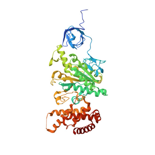
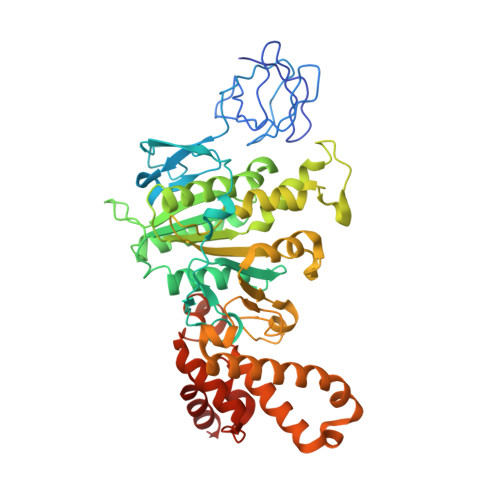
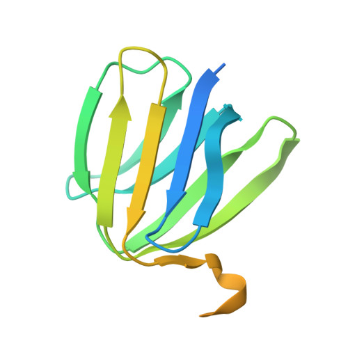
1H8E

(ADP.AlF4)2(ADP.SO4) bovine F1-ATPase (all three catalytic sites occupied)


Experimental Data Snapshot

  • Method: X-RAY DIFFRACTION
  • Resolution: 2.00 Å
  • R-Value Free: 0.264 
  • R-Value Work: 0.201 

Starting Model: experimental
View more details

wwPDB Validation   3D Report Full Report


Ligand Structure Quality Assessment 


This is version 1.3 of the entry. See complete history


Literature

Structure of Bovine Mitochondrial F1-ATPase with Nucleotide Bound to All Three Catalytic Sites: Implications for the Mechanism of Rotary Catalysis

Menz, R.I.Walker, J.E.Leslie, A.G.W.

(2001) Cell 106: 331

  • DOI: https://doi.org/10.1016/s0092-8674(01)00452-4
  • Primary Citation of Related Structures:  
    1H8E

  • PubMed Abstract: 

    The crystal structure of a novel aluminium fluoride inhibited form of bovine mitochondrial F(1)-ATPase has been determined at 2 A resolution. In contrast to all previously determined structures of the bovine enzyme, all three catalytic sites are occupied by nucleotide. The subunit that did not bind nucleotide in previous structures binds ADP and sulfate (mimicking phosphate), and adopts a "half-closed" conformation. This structure probably represents the posthydrolysis, pre-product release step on the catalytic pathway. A catalytic scheme for hydrolysis (and synthesis) at physiological rates and a mechanism for the ATP-driven rotation of the gamma subunit are proposed based on the crystal structures of the bovine enzyme.


  • Organizational Affiliation

    Medical Research Council Laboratory of Molecular Biology, Hills Road, Cambridge CB2 2QH, United Kingdom.


Macromolecules
Find similar proteins by:  (by identity cutoff)  |  3D Structure
Entity ID: 1
MoleculeChains Sequence LengthOrganismDetailsImage
BOVINE MITOCHONDRIAL F1-ATPASE
A, B, C
510Bos taurusMutation(s): 1 
UniProt
Find proteins for P19483 (Bos taurus)
Explore P19483 
Go to UniProtKB:  P19483
Entity Groups  
Sequence Clusters30% Identity50% Identity70% Identity90% Identity95% Identity100% Identity
UniProt GroupP19483
Sequence Annotations
Expand
  • Reference Sequence
Find similar proteins by:  (by identity cutoff)  |  3D Structure
Entity ID: 2
MoleculeChains Sequence LengthOrganismDetailsImage
BOVINE MITOCHONDRIAL F1-ATPASE
D, E, F
482Bos taurusMutation(s): 0 
UniProt
Find proteins for P00829 (Bos taurus)
Explore P00829 
Go to UniProtKB:  P00829
Entity Groups  
Sequence Clusters30% Identity50% Identity70% Identity90% Identity95% Identity100% Identity
UniProt GroupP00829
Sequence Annotations
Expand
  • Reference Sequence
Find similar proteins by:  (by identity cutoff)  |  3D Structure
Entity ID: 3
MoleculeChains Sequence LengthOrganismDetailsImage
BOVINE MITOCHONDRIAL F1-ATPASE272Bos taurusMutation(s): 0 
UniProt
Find proteins for P05631 (Bos taurus)
Explore P05631 
Go to UniProtKB:  P05631
Entity Groups  
Sequence Clusters30% Identity50% Identity70% Identity90% Identity95% Identity100% Identity
UniProt GroupP05631
Sequence Annotations
Expand
  • Reference Sequence
Find similar proteins by:  (by identity cutoff)  |  3D Structure
Entity ID: 4
MoleculeChains Sequence LengthOrganismDetailsImage
BOVINE MITOCHONDRIAL F1-ATPASE146Bos taurusMutation(s): 0 
UniProt
Find proteins for P05630 (Bos taurus)
Explore P05630 
Go to UniProtKB:  P05630
Entity Groups  
Sequence Clusters30% Identity50% Identity70% Identity90% Identity95% Identity100% Identity
UniProt GroupP05630
Sequence Annotations
Expand
  • Reference Sequence
Find similar proteins by:  (by identity cutoff)  |  3D Structure
Entity ID: 5
MoleculeChains Sequence LengthOrganismDetailsImage
BOVINE MITOCHONDRIAL F1-ATPASE50Bos taurusMutation(s): 0 
UniProt
Find proteins for P05632 (Bos taurus)
Explore P05632 
Go to UniProtKB:  P05632
Entity Groups  
Sequence Clusters30% Identity50% Identity70% Identity90% Identity95% Identity100% Identity
UniProt GroupP05632
Sequence Annotations
Expand
  • Reference Sequence
Small Molecules
Ligands 5 Unique
IDChains Name / Formula / InChI Key2D Diagram3D Interactions
ADP
Query on ADP

Download Ideal Coordinates CCD File 
J [auth A]
M [auth B]
Q [auth C]
T [auth D]
W [auth E]
J [auth A],
M [auth B],
Q [auth C],
T [auth D],
W [auth E],
Z [auth F]
ADENOSINE-5'-DIPHOSPHATE
C10 H15 N5 O10 P2
XTWYTFMLZFPYCI-KQYNXXCUSA-N
ALF
Query on ALF

Download Ideal Coordinates CCD File 
BA [auth F],
V [auth D]
TETRAFLUOROALUMINATE ION
Al F4
UYOMQIYKOOHAMK-UHFFFAOYSA-J
SO4
Query on SO4

Download Ideal Coordinates CCD File 
Y [auth E]SULFATE ION
O4 S
QAOWNCQODCNURD-UHFFFAOYSA-L
GOL
Query on GOL

Download Ideal Coordinates CCD File 
L [auth A],
O [auth B],
P [auth B],
S [auth C]
GLYCEROL
C3 H8 O3
PEDCQBHIVMGVHV-UHFFFAOYSA-N
MG
Query on MG

Download Ideal Coordinates CCD File 
AA [auth F]
K [auth A]
N [auth B]
R [auth C]
U [auth D]
AA [auth F],
K [auth A],
N [auth B],
R [auth C],
U [auth D],
X [auth E]
MAGNESIUM ION
Mg
JLVVSXFLKOJNIY-UHFFFAOYSA-N
Experimental Data & Validation

Experimental Data

  • Method: X-RAY DIFFRACTION
  • Resolution: 2.00 Å
  • R-Value Free: 0.264 
  • R-Value Work: 0.201 
  • Space Group: P 21 21 21
Unit Cell:
Length ( Å )Angle ( ˚ )
a = 267.7α = 90
b = 106.2β = 90
c = 138.3γ = 90
Software Package:
Software NamePurpose
REFMACrefinement
MOSFLMdata reduction
CCP4data scaling
AMoREphasing

Structure Validation

View Full Validation Report


Ligand Structure Quality Assessment 


Entry History 

Deposition Data

Revision History  (Full details and data files)

  • Version 1.0: 2001-08-10
    Type: Initial release
  • Version 1.1: 2011-05-08
    Changes: Version format compliance
  • Version 1.2: 2011-07-13
    Changes: Version format compliance
  • Version 1.3: 2023-12-13
    Changes: Data collection, Database references, Derived calculations, Other, Refinement description