1H5Q | pdb_00001h5q

Mannitol dehydrogenase from Agaricus bisporus


Experimental Data Snapshot

  • Method: X-RAY DIFFRACTION
  • Resolution: 1.50 Å
  • R-Value Free: 
    0.209 (Depositor), 0.218 (DCC) 
  • R-Value Work: 
    0.193 (Depositor) 
  • R-Value Observed: 
    0.193 (Depositor) 

Starting Model: experimental
View more details

wwPDB Validation 3D Report Full Report

Validation slider image for 1H5Q

Ligand Structure Quality Assessment 


This is version 1.3 of the entry. See complete history

Literature

The Crystallographic Structure of the Mannitol 2-Dehydrogenase Nadp+ Binary Complex from Agaricus Bisporus

Horer, S.Stoop, J.Mooibroek, H.Baumann, U.Sassoon, J.

(2001) J Biological Chem 276: 27555

  • DOI: https://doi.org/10.1074/jbc.M102850200
  • Primary Citation Related Structures: 
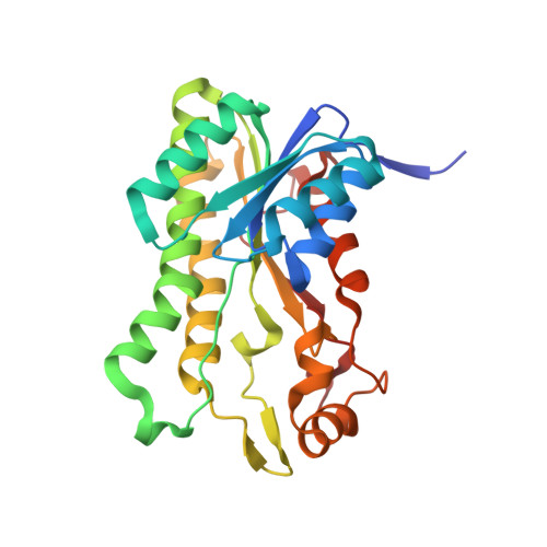
    1H5Q

  • PubMed Abstract: 

    Mannitol, an acyclic six-carbon polyol, is one of the most abundant sugar alcohols occurring in nature. In the button mushroom, Agaricus bisporus, it is synthesized from fructose by the enzyme mannitol 2-dehydrogenase (MtDH; EC ) using NADPH as a cofactor. Mannitol serves as the main storage carbon (up to 50% of the fruit body dry weight) and plays a critical role in growth, fruit body development, osmoregulation, and salt tolerance. Furthermore, mannitol dehydrogenases are being evaluated for commercial mannitol production as alternatives to the less efficient chemical reduction of fructose. Given the importance of mannitol metabolism and mannitol dehydrogenases, MtDH was cloned into the pET28 expression system and overexpressed in Escherichia coli. Kinetic and physicochemical properties of the recombinant enzyme are indistinguishable from the natural enzyme. The crystal structure of its binary complex with NADP was solved at 1.5-A resolution and refined to an R value of 19.3%. It shows MtDH to be a tetramer and a member of the short chain dehydrogenase/reductase family of enzymes. The catalytic residues forming the so-called catalytic triad can be assigned to Ser(149), Tyr(169), and Lys(173).


  • Organizational Affiliation
    • Department of Chemistry and Biochemistry, University of Berne, Freiestrasse 3, 3012 Berne, Switzerland.

Macromolecule Content 

  • Total Structure Weight: 348.13 kDa 
  • Atom Count: 27,197 
  • Modeled Residue Count: 3,120 
  • Deposited Residue Count: 3,180 
  • Unique protein chains: 1

Macromolecules

Find similar proteins by:|  3D Structure
Entity ID: 1
MoleculeChains  Sequence LengthOrganismDetailsImage
NADP-DEPENDENT MANNITOL DEHYDROGENASE
A, B, C, D, E
A, B, C, D, E, F, G, H, I, J, K, L
265Agaricus bisporusMutation(s): 0 
EC: 1.1.1.138
UniProt
Find proteins for O93868 (Agaricus bisporus)
Explore O93868 
Go to UniProtKB:  O93868
Entity Groups
Sequence Clusters30% Identity50% Identity70% Identity90% Identity95% Identity100% Identity
UniProt GroupO93868
Sequence Annotations
Expand
Reference Sequence

Small Molecules

Ligands 2 Unique
IDChains Name / Formula / InChI Key2D Diagram3D Interactions
NAP

Query on NAP


Download:Ideal Coordinates CCD File
AA [auth J]
CA [auth K]
DA [auth L]
M [auth A]
O [auth B]
AA [auth J],
CA [auth K],
DA [auth L],
M [auth A],
O [auth B],
Q [auth C],
R [auth D],
S [auth E],
U [auth F],
W [auth G],
X [auth H],
Y [auth I]
NADP NICOTINAMIDE-ADENINE-DINUCLEOTIDE PHOSPHATE
C21 H28 N7 O17 P3
XJLXINKUBYWONI-NNYOXOHSSA-N
NI

Query on NI


Download:Ideal Coordinates CCD File
BA [auth J]
N [auth A]
P [auth B]
T [auth E]
V [auth F]
BA [auth J],
N [auth A],
P [auth B],
T [auth E],
V [auth F],
Z [auth I]
NICKEL (II) ION
Ni
VEQPNABPJHWNSG-UHFFFAOYSA-N

Experimental Data & Validation

Experimental Data

  • Method: X-RAY DIFFRACTION
  • Resolution: 1.50 Å
  • R-Value Free:  0.209 (Depositor), 0.218 (DCC) 
  • R-Value Work:  0.193 (Depositor) 
  • R-Value Observed: 0.193 (Depositor) 
Space Group: C 1 2 1
Unit Cell:
Length ( Å )Angle ( ˚ )
a = 227.25α = 90
b = 124.85β = 118.54
c = 132.69γ = 90
Software Package:
Software NamePurpose
REFMACrefinement
MOSFLMdata reduction
SCALAdata scaling
CNSphasing

Structure Validation

View Full Validation Report


Ligand Structure Quality Assessment 


Entry History 

Deposition Data

Revision History  (Full details and data files)

  • Version 1.0: 2001-06-21
    Type: Initial release
  • Version 1.1: 2011-05-08
    Changes: Version format compliance
  • Version 1.2: 2011-07-13
    Changes: Version format compliance
  • Version 1.3: 2023-12-13
    Changes: Data collection, Database references, Derived calculations, Refinement description