1H3P | pdb_00001h3p

STRUCTURAL CHARACTERISATION OF A MONOCLONAL ANTIBODY SPECIFIC FOR THE PRES1 REGION OF THE HEPATITIS B VIRUS


Experimental Data Snapshot

  • Method: X-RAY DIFFRACTION
  • Resolution: 2.60 Å
  • R-Value Free: 
    0.291 (Depositor), 0.278 (DCC) 
  • R-Value Work: 
    0.213 (Depositor), 0.203 (DCC) 
  • R-Value Observed: 
    0.213 (Depositor) 

Starting Model: experimental
View more details

wwPDB Validation 3D Report Full Report

Validation slider image for 1H3P

This is version 1.4 of the entry. See complete history

Literature

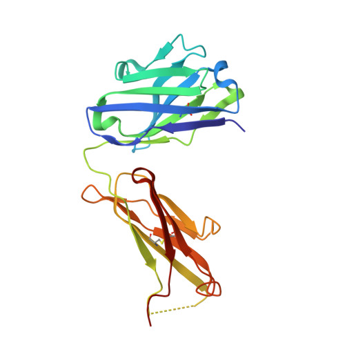
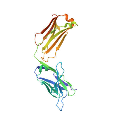
Structural and Functional Characterisation of a Monoclonal Antibody Specific for the Pres1 Region of Hepatitis B Virus

Pizarro, J.C.Vulliez-Le-Normand, B.Riottot, M.M.Budkowska, A.Bentley, G.A.

(2001) FEBS Lett 509: 463

  • DOI: https://doi.org/10.1016/s0014-5793(01)03190-8
  • Primary Citation Related Structures: 
    1H3P

  • PubMed Abstract: 

    The monoclonal antibody 5a19, raised against the ay serotype of hepatitis B virus, binds to the segment of the preS1 region comprising residues 37-43, which is implicated in attachment of the virus to hepatocytes. The dissociation constant, derived from kinetic studies using surface plasmon resonance techniques, is in the low nanomolar range. The nucleotide sequence of the variable domains has been determined and the corresponding germ-line genes have been identified. The three-dimensional structure of the Fab fragment has been determined by X-ray crystallography to 2.6 A resolution.


  • Organizational Affiliation
    • Unité d' Immunologie Structurale, Institut Pasteur, Paris, France.

Macromolecule Content 

  • Total Structure Weight: 49.81 kDa 
  • Atom Count: 3,450 
  • Modeled Residue Count: 435 
  • Deposited Residue Count: 459 
  • Unique protein chains: 2

Macromolecules

Find similar proteins by:|  3D Structure
Entity ID: 1
MoleculeChains  Sequence LengthOrganismDetailsImage
ANTIBODY FAB FRAGMENTA [auth H]219Mus musculusMutation(s): 0 
UniProt
Find proteins for Q65ZL8 (Mus musculus)
Explore Q65ZL8 
Go to UniProtKB:  Q65ZL8
Entity Groups
Sequence Clusters30% Identity50% Identity70% Identity90% Identity95% Identity100% Identity
UniProt GroupQ65ZL8
Sequence Annotations
Expand
Reference Sequence
Find similar proteins by:|  3D Structure
Entity ID: 2
MoleculeChains  Sequence LengthOrganismDetailsImage
ANTIBODY FAB FRAGMENTB [auth L]240Mus musculusMutation(s): 0 
UniProt
Find proteins for Q52L64 (Mus musculus)
Explore Q52L64 
Go to UniProtKB:  Q52L64
Entity Groups
Sequence Clusters30% Identity50% Identity70% Identity90% Identity95% Identity100% Identity
UniProt GroupQ52L64
Sequence Annotations
Expand
Reference Sequence

Experimental Data & Validation

Experimental Data

  • Method: X-RAY DIFFRACTION
  • Resolution: 2.60 Å
  • R-Value Free:  0.291 (Depositor), 0.278 (DCC) 
  • R-Value Work:  0.213 (Depositor), 0.203 (DCC) 
  • R-Value Observed: 0.213 (Depositor) 
Space Group: P 21 21 21
Unit Cell:
Length ( Å )Angle ( ˚ )
a = 47.01α = 90
b = 88.46β = 90
c = 111.67γ = 90
Software Package:
Software NamePurpose
X-PLORrefinement
DENZOdata reduction
SCALEPACKdata scaling
AMoREphasing

Structure Validation

View Full Validation Report


Entry History 

Deposition Data

Revision History  (Full details and data files)

  • Version 1.0: 2002-09-19
    Type: Initial release
  • Version 1.1: 2011-05-08
    Changes: Version format compliance
  • Version 1.2: 2011-07-13
    Changes: Version format compliance
  • Version 1.3: 2023-12-13
    Changes: Data collection, Database references, Refinement description
  • Version 1.4: 2024-10-09
    Changes: Structure summary