Structures of Phosphate and Trivanadate Complexes of Bacillus Stearothermophilus Phosphatase Phoe: Structural and Functional Analysis in the Cofactor-Dependent Phosphoglycerate Mutase Superfamily
Rigden, D.J., Littlejohn, J.E., Henderson, K., Jedrzejas, M.J.(2003) J Mol Biology 325: 411
- PubMed: 12498792 Search on PubMed
- DOI: https://doi.org/10.1016/s0022-2836(02)01229-9
- Primary Citation Related Structures:
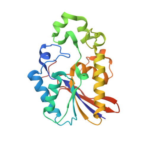
1H2E, 1H2F - PubMed Abstract:
Bacillus stearothermophilus phosphatase PhoE is a member of the cofactor-dependent phosphoglycerate mutase superfamily possessing broad specificity phosphatase activity. Its previous structural determination in complex with glycerol revealed probable bases for its efficient hydrolysis of both large, hydrophobic, and smaller, hydrophilic substrates. Here we report two further structures of PhoE complexes, to higher resolution of diffraction, which yield a better and thorough understanding of its catalytic mechanism. The environment of the phosphate ion in the catalytic site of the first complex strongly suggests an acid-base catalytic function for Glu83. It also reveals how the C-terminal tail ordering is linked to enzyme activation on phosphate binding by a different mechanism to that seen in Escherichia coli phosphoglycerate mutase. The second complex structure with an unusual doubly covalently bound trivanadate shows how covalent modification of the phosphorylable His10 is accompanied by small structural changes, presumably to catalytic advantage. When compared with structures of related proteins in the cofactor-dependent phosphoglycerate mutase superfamily, an additional phosphate ligand, Gln22, is observed in PhoE. Functional constraints lead to the corresponding residue being conserved as Gly in fructose-2,6-bisphosphatases and Thr/Ser/Cys in phosphoglycerate mutases. A number of sequence annotation errors in databases are highlighted by this analysis. B. stearothermophilus PhoE is evolutionarily related to a group of enzymes primarily present in Gram-positive bacilli. Even within this group substrate specificity is clearly variable highlighting the difficulties of computational functional annotation in the cofactor-dependent phosphoglycerate mutase superfamily.
- National Centre of Genetic Resources and Biotechnology, Cenargen/Embrapa, SAIN Parque Rural, Final W5, Asa Norte, 70770-900 Brasília, Brazil.
Organizational Affiliation: