1GZU | pdb_00001gzu

Crystal Structure of Human Nicotinamide Mononucleotide Adenylyltransferase in Complex with NMN


Experimental Data Snapshot

  • Method: X-RAY DIFFRACTION
  • Resolution: 2.90 Å
  • R-Value Free: 
    0.286 (Depositor) 
  • R-Value Work: 
    0.246 (Depositor) 
  • R-Value Observed: 
    0.248 (Depositor) 

wwPDB Validation 3D Report Full Report

Validation slider image for 1GZU

This is version 1.4 of the entry. See complete history

Literature

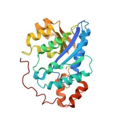
Crystal structure of human nicotinamide mononucleotide adenylyltransferase in complex with NMN.

Werner, E.Ziegler, M.Lerner, F.Schweiger, M.Heinemann, U.

(2002) FEBS Lett 516: 239-244

  • DOI: https://doi.org/10.1016/s0014-5793(02)02556-5
  • Primary Citation Related Structures: 
    1GZU

  • PubMed Abstract: 

    The final step in the biosynthesis of nicotinamide-adenine dinucleotide, a major coenzyme in cellular redox reactions and involved in intracellular signaling, is catalyzed by the enzyme nicotinamide mononucleotide adenylyltransferase (NMNAT). The X-ray structure of human NMNAT in complex with nicotinamide mononucleotide was solved by the single-wavelength anomalous dispersion method at a resolution of 2.9 A. Human NMNAT is a symmetric hexamer whose subunit is formed by a large six-stranded parallel beta-sheet with helices on both sides. Human NMNAT displays a different oligomerization compared to the archaeal enzyme. The protein-nicotinamide mononucleotide interaction pattern provides insight into ligand binding in the human enzyme.


  • Organizational Affiliation
    • Crystallography Group, Max Delbrück Center for Molecular Medicine, Robert-Rössle-Str. 10, D-13092, Berlin, Germany. ewe@mdc-berlin.de

Macromolecule Content 

  • Total Structure Weight: 101.34 kDa 
  • Atom Count: 5,544 
  • Modeled Residue Count: 684 
  • Deposited Residue Count: 870 
  • Unique protein chains: 1

Macromolecules

Find similar proteins by:|  3D Structure
Entity ID: 1
MoleculeChains  Sequence LengthOrganismDetailsImage
NICOTINAMIDE MONONUCLEOTIDE ADENYLYL TRANSFERASE
A, B, C
290Homo sapiensMutation(s): 0 
EC: 2.7.7.1 (PDB Primary Data), 2.7.7.18 (UniProt)
UniProt & NIH Common Fund Data Resources
Find proteins for Q9HAN9 (Homo sapiens)
Explore Q9HAN9 
Go to UniProtKB:  Q9HAN9
PHAROS:  Q9HAN9
GTEx:  ENSG00000173614 
Entity Groups
Sequence Clusters30% Identity50% Identity70% Identity90% Identity95% Identity100% Identity
UniProt GroupQ9HAN9
Sequence Annotations
Expand
Reference Sequence

Small Molecules

Modified Residues  1 Unique
IDChains TypeFormula2D DiagramParent
MSE
Query on MSE
A, B, C
L-PEPTIDE LINKINGC5 H11 N O2 SeMET

Experimental Data & Validation

Experimental Data

  • Method: X-RAY DIFFRACTION
  • Resolution: 2.90 Å
  • R-Value Free:  0.286 (Depositor) 
  • R-Value Work:  0.246 (Depositor) 
  • R-Value Observed: 0.248 (Depositor) 
Space Group: C 2 2 21
Unit Cell:
Length ( Å )Angle ( ˚ )
a = 140.8α = 90
b = 235.7β = 90
c = 89γ = 90
Software Package:
Software NamePurpose
REFMACrefinement
DENZOdata reduction
SCALEPACKdata scaling
SOLVEphasing

Structure Validation

View Full Validation Report


Entry History 

Deposition Data

Revision History  (Full details and data files)

  • Version 1.0: 2002-07-05
    Type: Initial release
  • Version 1.1: 2011-05-08
    Changes: Version format compliance
  • Version 1.2: 2011-07-13
    Changes: Version format compliance
  • Version 1.3: 2018-06-13
    Changes: Advisory, Data collection, Database references, Refinement description, Structure summary
  • Version 1.4: 2024-11-13
    Changes: Advisory, Data collection, Database references, Derived calculations, Refinement description, Structure summary