1GZQ

CD1b in complex with Phophatidylinositol


Experimental Data Snapshot

  • Method: X-RAY DIFFRACTION
  • Resolution: 2.26 Å
  • R-Value Free: 0.237 
  • R-Value Work: 0.203 
  • R-Value Observed: 0.203 

wwPDB Validation   3D Report Full Report


Ligand Structure Quality Assessment 


This is version 1.4 of the entry. See complete history


Literature

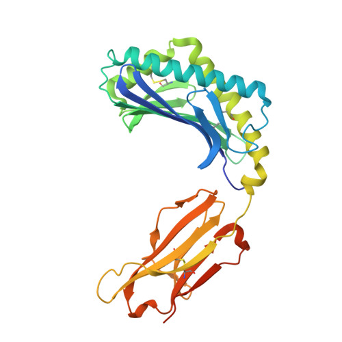
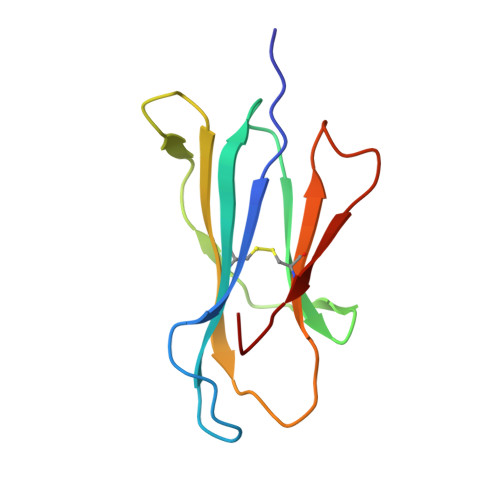
Structure of Human Cd1B with Bound Ligands at 2.3 A, a Maze for Alkyl Chains

Gadola, S.D.Zaccai, N.R.Harlos, K.Shepherd, D.Castro-Palomino, J.C.Ritter, G.Schmidt, R.R.Jones, E.Y.Cerundolo, V.

(2002) Nat Immunol 3: 721

  • DOI: https://doi.org/10.1038/ni821
  • Primary Citation of Related Structures:  
    1GZP, 1GZQ

  • PubMed Abstract: 

    The human genome encodes five nonpolymorphic major histocompatibility complex class I-like glycoproteins, CD1a to CD1e, that present lipid antigens for specific recognition by T lymphocytes. Using single alkyl chain detergents, we developed a protocol to generate recombinant human CD1b-lipid complexes. We present here the crystal structures of CD1b in complex with either phosphatidylinositol or ganglioside GM2 at 2.3 A and 2.8 A resolutions, respectively. The antigen-binding groove houses four interlinked hydrophobic channels that are occupied by the alkyl chains of the glycolipid plus two detergent molecules. A distinct exit beneath the alpha 2 helix further contributes to the plasticity of the binding groove. These structures reveal the mechanism by which two alkyl chain lipids bind to CD1b, and how CD1b can adapt to ligands of different alkyl chain length. They also suggest how very long alkyl chains, such as those of mycolic acid, could be fully contained within the binding groove. These results extend the spectrum of potential CD1b ligands by revealing that, in addition to two alkyl chain lipids, mono-alkyl and triple-alkyl chain lipids can be accommodated in the binding groove.


  • Organizational Affiliation

    Cancer Research UK Tumour Immunology Group, The Weatherall Institute of Molecular Medicine, Nuffield Department of Medicine, University of Oxford, Oxford OX3 9DS, UK. stephan.gadola@inselch


Macromolecules
Find similar proteins by:  (by identity cutoff)  |  3D Structure
Entity ID: 1
MoleculeChains Sequence LengthOrganismDetailsImage
T-CELL SURFACE GLYCOPROTEIN CD1B300Homo sapiensMutation(s): 0 
UniProt & NIH Common Fund Data Resources
Find proteins for P29016 (Homo sapiens)
Explore P29016 
Go to UniProtKB:  P29016
PHAROS:  P29016
GTEx:  ENSG00000158485 
Entity Groups  
Sequence Clusters30% Identity50% Identity70% Identity90% Identity95% Identity100% Identity
UniProt GroupP29016
Sequence Annotations
Expand
  • Reference Sequence
Find similar proteins by:  (by identity cutoff)  |  3D Structure
Entity ID: 2
MoleculeChains Sequence LengthOrganismDetailsImage
B2-MICROGLOBULIN100Homo sapiensMutation(s): 0 
UniProt & NIH Common Fund Data Resources
Find proteins for P61769 (Homo sapiens)
Explore P61769 
Go to UniProtKB:  P61769
PHAROS:  P61769
GTEx:  ENSG00000166710 
Entity Groups  
Sequence Clusters30% Identity50% Identity70% Identity90% Identity95% Identity100% Identity
UniProt GroupP61769
Sequence Annotations
Expand
  • Reference Sequence
Small Molecules
Ligands 4 Unique
IDChains Name / Formula / InChI Key2D Diagram3D Interactions
PII
Query on PII

Download Ideal Coordinates CCD File 
C [auth A]2-[(HYDROXY{[(2R,3R,5S,6R)-2,3,4,5,6-PENTAHYDROXYCYCLOHEXYL]OXY}PHOSPHORYL)OXY]-1-[(PALMITOYLOXY)METHYL]ETHYL HEPTADECANOATE
C42 H81 O13 P
QVAGJHIIXLLVCI-KSGPIMHQSA-N
TWT
Query on TWT

Download Ideal Coordinates CCD File 
H [auth A]DOCOSANE
C22 H46
HOWGUJZVBDQJKV-UHFFFAOYSA-N
D12
Query on D12

Download Ideal Coordinates CCD File 
G [auth A]DODECANE
C12 H26
SNRUBQQJIBEYMU-UHFFFAOYSA-N
NO3
Query on NO3

Download Ideal Coordinates CCD File 
D [auth A],
E [auth A],
F [auth A]
NITRATE ION
N O3
NHNBFGGVMKEFGY-UHFFFAOYSA-N
Experimental Data & Validation

Experimental Data

  • Method: X-RAY DIFFRACTION
  • Resolution: 2.26 Å
  • R-Value Free: 0.237 
  • R-Value Work: 0.203 
  • R-Value Observed: 0.203 
  • Space Group: C 2 2 21
Unit Cell:
Length ( Å )Angle ( ˚ )
a = 87.881α = 90
b = 176.998β = 90
c = 75.277γ = 90
Software Package:
Software NamePurpose
CNSrefinement
DENZOdata reduction
SCALEPACKdata scaling
AMoREphasing

Structure Validation

View Full Validation Report


Ligand Structure Quality Assessment 


Entry History 

Deposition Data

Revision History  (Full details and data files)

  • Version 1.0: 2002-07-31
    Type: Initial release
  • Version 1.1: 2011-05-08
    Changes: Version format compliance
  • Version 1.2: 2011-07-13
    Changes: Version format compliance
  • Version 1.3: 2017-08-09
    Changes: Data collection, Source and taxonomy
  • Version 1.4: 2019-05-08
    Changes: Data collection, Experimental preparation