1GZN | pdb_00001gzn

Structure of PKB kinase domain


Experimental Data Snapshot

  • Method: X-RAY DIFFRACTION
  • Resolution: 2.50 Å
  • R-Value Free: 
    0.319 (Depositor), 0.301 (DCC) 
  • R-Value Work: 
    0.268 (Depositor), 0.249 (DCC) 
  • R-Value Observed: 
    0.268 (Depositor) 

Starting Model: experimental
View more details

wwPDB Validation 3D Report Full Report

Validation slider image for 1GZN

This is version 1.3 of the entry. See complete history

Literature

Molecular Mechanism for the Regulation of Protein Kinase B/Akt by Hydrophobic Motif Phosphorylation

Yang, J.Cron, P.Thompson, V.Good, V.Hess, D.Hemmings, B.A.Barford, D.

(2002) Mol Cell 9: 1227

  • DOI: https://doi.org/10.1016/s1097-2765(02)00550-6
  • Primary Citation Related Structures: 
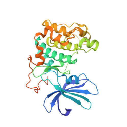
    1GZK, 1GZN, 1GZO

  • PubMed Abstract: 

    Protein kinase B/Akt plays crucial roles in promoting cell survival and mediating insulin responses. The enzyme is stimulated by phosphorylation at two regulatory sites: Thr 309 of the activation segment and Ser 474 of the hydrophobic motif, a conserved feature of many AGC kinases. Analysis of the crystal structures of the unphosphorylated and Thr 309 phosphorylated states of the PKB kinase domain provides a molecular explanation for regulation by Ser 474 phosphorylation. Activation by Ser 474 phosphorylation occurs via a disorder to order transition of the alphaC helix with concomitant restructuring of the activation segment and reconfiguration of the kinase bilobal structure. These conformational changes are mediated by a phosphorylation-promoted interaction of the hydrophobic motif with a channel on the N-terminal lobe induced by the ordered alphaC helix and are mimicked by peptides corresponding to the hydrophobic motif of PKB and potently by the hydrophobic motif of PRK2.


  • Organizational Affiliation
    • Section of Structural Biology, Institute of Cancer Research, Chester Beatty Laboratories, 237 Fulham Road, London SW3 6JB, UK.

Macromolecule Content 

  • Total Structure Weight: 38.88 kDa 
  • Atom Count: 2,445 
  • Modeled Residue Count: 272 
  • Deposited Residue Count: 335 
  • Unique protein chains: 1

Macromolecules

Find similar proteins by:|  3D Structure
Entity ID: 1
MoleculeChains  Sequence LengthOrganismDetailsImage
RAC-BETA SERINE/THREONINE PROTEIN KINASE335Homo sapiensMutation(s): 0 
EC: 2.7.1 (PDB Primary Data), 2.7.11.1 (UniProt)
UniProt & NIH Common Fund Data Resources
Find proteins for P31751 (Homo sapiens)
Explore P31751 
Go to UniProtKB:  P31751
PHAROS:  P31751
GTEx:  ENSG00000105221 
Entity Groups
Sequence Clusters30% Identity50% Identity70% Identity90% Identity95% Identity100% Identity
UniProt GroupP31751
Sequence Annotations
Expand
Reference Sequence

Experimental Data & Validation

Experimental Data

  • Method: X-RAY DIFFRACTION
  • Resolution: 2.50 Å
  • R-Value Free:  0.319 (Depositor), 0.301 (DCC) 
  • R-Value Work:  0.268 (Depositor), 0.249 (DCC) 
  • R-Value Observed: 0.268 (Depositor) 
Space Group: P 41 21 2
Unit Cell:
Length ( Å )Angle ( ˚ )
a = 149.523α = 90
b = 149.523β = 90
c = 39.055γ = 90
Software Package:
Software NamePurpose
CNSrefinement
DENZOdata reduction
SCALEPACKdata scaling
CNSphasing

Structure Validation

View Full Validation Report


Entry History 

Deposition Data

Revision History  (Full details and data files)

  • Version 1.0: 2003-05-22
    Type: Initial release
  • Version 1.1: 2011-05-08
    Changes: Version format compliance
  • Version 1.2: 2011-07-13
    Changes: Version format compliance
  • Version 1.3: 2023-12-13
    Changes: Data collection, Database references, Other, Refinement description