1GYW | pdb_00001gyw

Gamma-adaptin appendage domain from clathrin adaptor AP1 A753D mutant


Experimental Data Snapshot

  • Method: X-RAY DIFFRACTION
  • Resolution: 2.40 Å
  • R-Value Free: 
    0.269 (Depositor), 0.268 (DCC) 
  • R-Value Work: 
    0.190 (Depositor), 0.200 (DCC) 

Starting Model: experimental
View more details

wwPDB Validation 3D Report Full Report

Validation slider image for 1GYW

This is version 1.6 of the entry. See complete history

Literature

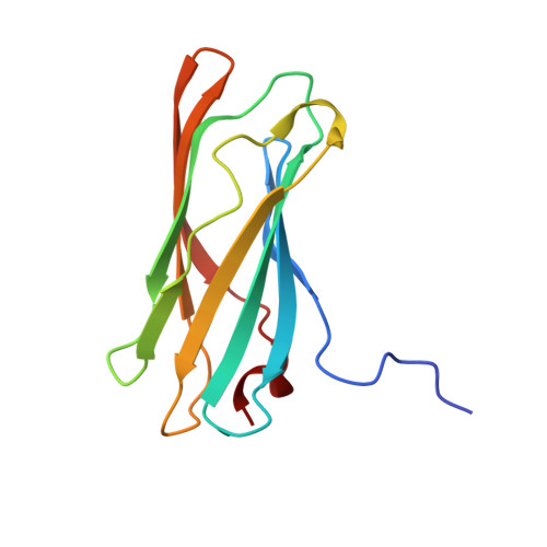
Gamma-adaptin appendage domain: structure and binding site for Eps15 and gamma-synergin.

Kent, H.M.McMahon, H.T.Evans, P.R.Benmerah, A.Owen, D.J.

(2002) Structure 10: 1139-1148

  • DOI: https://doi.org/10.1016/s0969-2126(02)00801-8
  • Primary Citation Related Structures: 
    1GYU, 1GYV, 1GYW

  • PubMed Abstract: 

    The AP1 complex is one of a family of heterotetrameric clathrin-adaptor complexes involved in vesicular trafficking between the Golgi and endosomes. The complex has two large subunits, gamma and beta1, which can be divided into trunk, hinge, and appendage domains. The 1.8 A resolution structure of the gamma appendage is presented. The binding site for the known gamma appendage ligand gamma-synergin is mapped through creation of point mutations designed on the basis of the structure. We also show that Eps15, a protein believed to be involved in vesicle formation at the plasma membrane, is also a ligand of gamma appendage and binds to the same site as gamma-synergin. This observation explains the demonstrated brefeldinA (BFA)-sensitive colocalization of Eps15 and AP1 at the Golgi complex.


  • Organizational Affiliation
    • MRC Laboratory of Molecular Biology, Cambridge, United Kingdom.

Macromolecule Content 

  • Total Structure Weight: 28.55 kDa 
  • Atom Count: 2,172 
  • Modeled Residue Count: 254 
  • Deposited Residue Count: 256 
  • Unique protein chains: 1

Macromolecules

Find similar proteins by:|  3D Structure
Entity ID: 1
MoleculeChains  Sequence LengthOrganismDetailsImage
ADAPTER-RELATED PROTEIN COMPLEX 1 GAMMA 1 SUBUNIT
A, B
128Mus musculusMutation(s): 1 
UniProt & NIH Common Fund Data Resources
Find proteins for P22892 (Mus musculus)
Explore P22892 
Go to UniProtKB:  P22892
IMPC:  MGI:101919
Entity Groups
Sequence Clusters30% Identity50% Identity70% Identity90% Identity95% Identity100% Identity
UniProt GroupP22892
Sequence Annotations
Expand
Reference Sequence

Small Molecules

Ligands 1 Unique
IDChains Name / Formula / InChI Key2D Diagram3D Interactions
CL

Query on CL


Download:Ideal Coordinates CCD File
C [auth A]CHLORIDE ION
Cl
VEXZGXHMUGYJMC-UHFFFAOYSA-M

Experimental Data & Validation

Experimental Data

  • Method: X-RAY DIFFRACTION
  • Resolution: 2.40 Å
  • R-Value Free:  0.269 (Depositor), 0.268 (DCC) 
  • R-Value Work:  0.190 (Depositor), 0.200 (DCC) 
Space Group: P 43 21 2
Unit Cell:
Length ( Å )Angle ( ˚ )
a = 62.155α = 90
b = 62.155β = 90
c = 148.903γ = 90
Software Package:
Software NamePurpose
REFMACrefinement
MOSFLMdata reduction
SCALAdata scaling
AMoREphasing

Structure Validation

View Full Validation Report


Entry History 

Deposition Data

Revision History  (Full details and data files)

  • Version 1.0: 2002-08-22
    Type: Initial release
  • Version 1.1: 2011-05-08
    Changes: Version format compliance
  • Version 1.2: 2011-07-13
    Changes: Version format compliance
  • Version 1.3: 2018-02-28
    Changes: Database references, Source and taxonomy
  • Version 1.4: 2019-03-06
    Changes: Data collection, Experimental preparation
  • Version 1.5: 2019-10-09
    Changes: Data collection, Database references, Other
  • Version 1.6: 2023-12-13
    Changes: Data collection, Database references, Refinement description